Summary information and primary citation
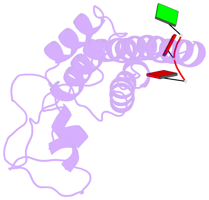
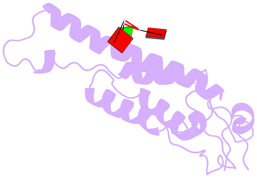
- PDB-id
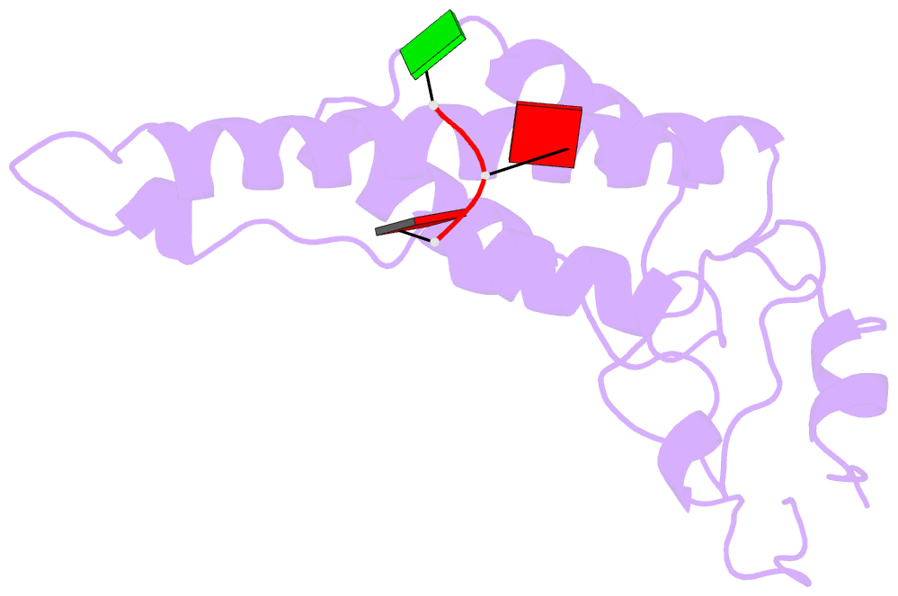
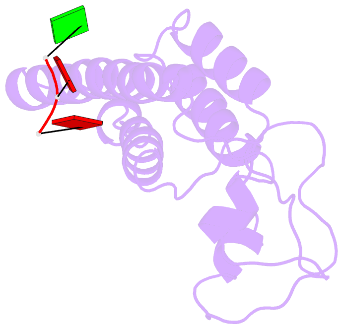
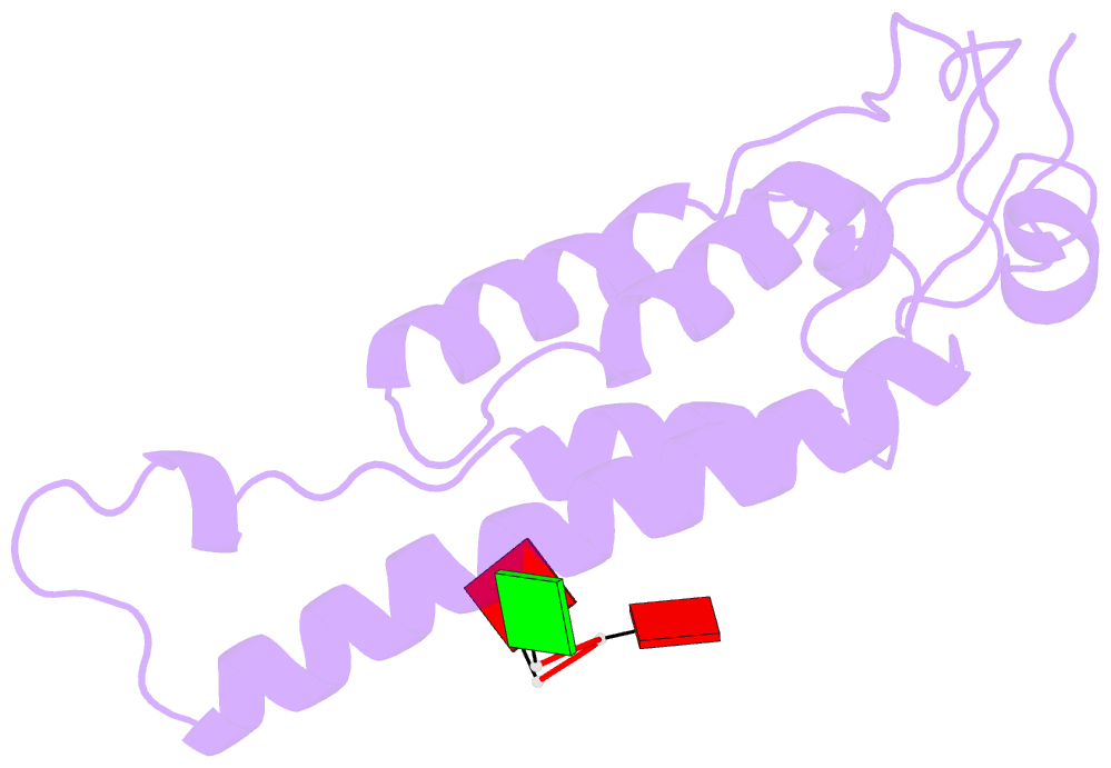
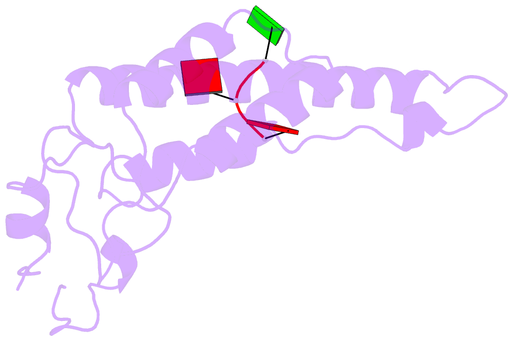
- 7q22; DSSR-derived features in text and JSON formats
- Class
- virus
- Method
- cryo-EM (6.3 Å)
- Summary
- Cryo idpc-stem structure recorded with csa 2.0
- Reference
- Lazic I, Wirix M, Leidl ML, de Haas F, Mann D, Beckers M, Pechnikova EV, Muller-Caspary K, Egoavil R, Bosch EGT, Sachse C (2022): "Single-particle cryo-EM structures from iDPC-STEM at near-atomic resolution." Nat.Methods, 19, 1126-1136. doi: 10.1038/s41592-022-01586-0.
- Abstract
- In electron cryomicroscopy (cryo-EM), molecular images of vitrified biological samples are obtained by conventional transmission microscopy (CTEM) using large underfocuses and subsequently computationally combined into a high-resolution three-dimensional structure. Here, we apply scanning transmission electron microscopy (STEM) using the integrated differential phase contrast mode also known as iDPC-STEM to two cryo-EM test specimens, keyhole limpet hemocyanin (KLH) and tobacco mosaic virus (TMV). The micrographs show complete contrast transfer to high resolution and enable the cryo-EM structure determination for KLH at 6.5 Å resolution, as well as for TMV at 3.5 Å resolution using single-particle reconstruction methods, which share identical features with maps obtained by CTEM of a previously acquired same-sized TMV data set. These data show that STEM imaging in general, and in particular the iDPC-STEM approach, can be applied to vitrified single-particle specimens to determine near-atomic resolution cryo-EM structures of biological macromolecules.