Summary information and primary citation
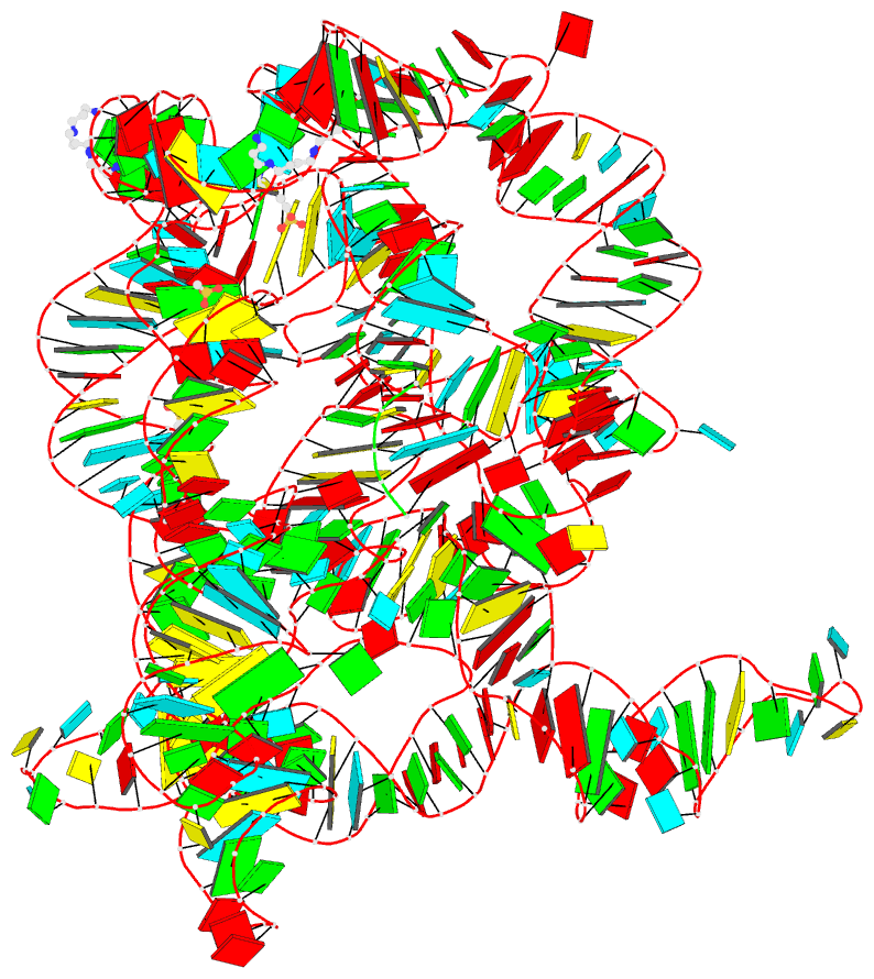
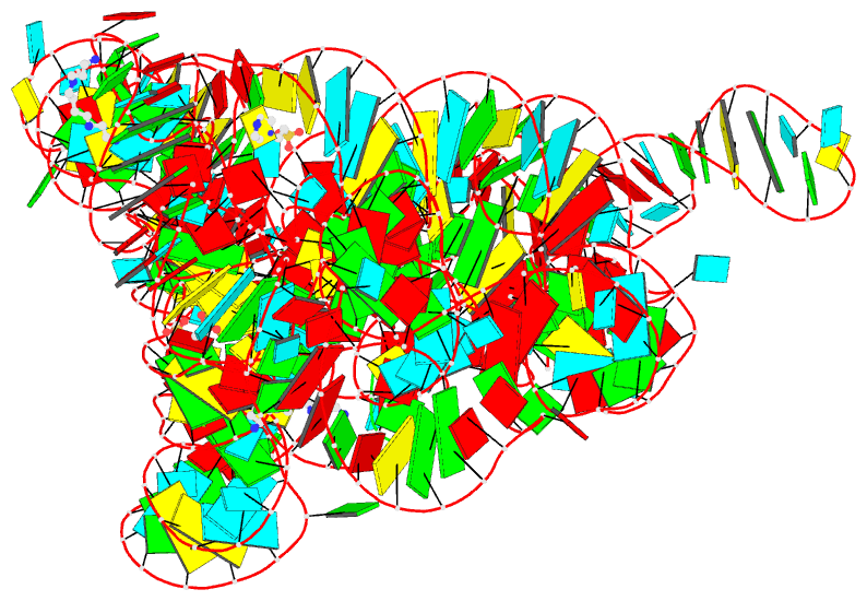
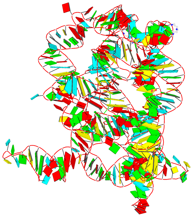
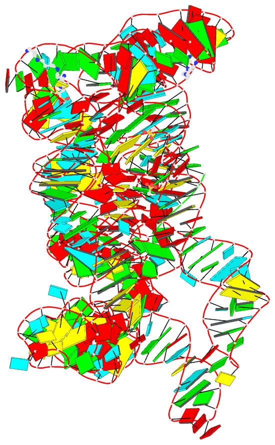
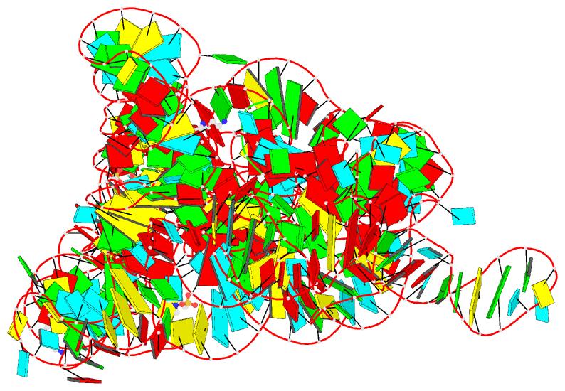
- PDB-id
- 6t3k; DSSR-derived features in text and JSON formats
- Class
- RNA
- Method
- X-ray (3.44 Å)
- Summary
- Structure of oceanobacillus iheyensis group ii intron g-mutant (c289g-c358g-g385c) in the presence of k+, mg2+ and 5'-exon
- Reference
- Manigrasso J, Chillon I, Genna V, Vidossich P, Somarowthu S, Pyle AM, De Vivo M, Marcia M (2020): "Visualizing group II intron dynamics between the first and second steps of splicing." Nat Commun, 11, 2837. doi: 10.1038/s41467-020-16741-4.
- Abstract
- Group II introns are ubiquitous self-splicing ribozymes and retrotransposable elements evolutionarily and chemically related to the eukaryotic spliceosome, with potential applications as gene-editing tools. Recent biochemical and structural data have captured the intron in multiple conformations at different stages of catalysis. Here, we employ enzymatic assays, X-ray crystallography, and molecular simulations to resolve the spatiotemporal location and function of conformational changes occurring between the first and the second step of splicing. We show that the first residue of the highly-conserved catalytic triad is protonated upon 5'-splice-site scission, promoting a reversible structural rearrangement of the active site (toggling). Protonation and active site dynamics induced by the first step of splicing facilitate the progression to the second step. Our insights into the mechanism of group II intron splicing parallels functional data on the spliceosome, thus reinforcing the notion that these evolutionarily-related molecular machines share the same enzymatic strategy.