Summary information and primary citation
- PDB-id
- 5y88; DSSR-derived features in text and JSON formats
- Class
- splicing
- Method
- cryo-EM (3.7 Å)
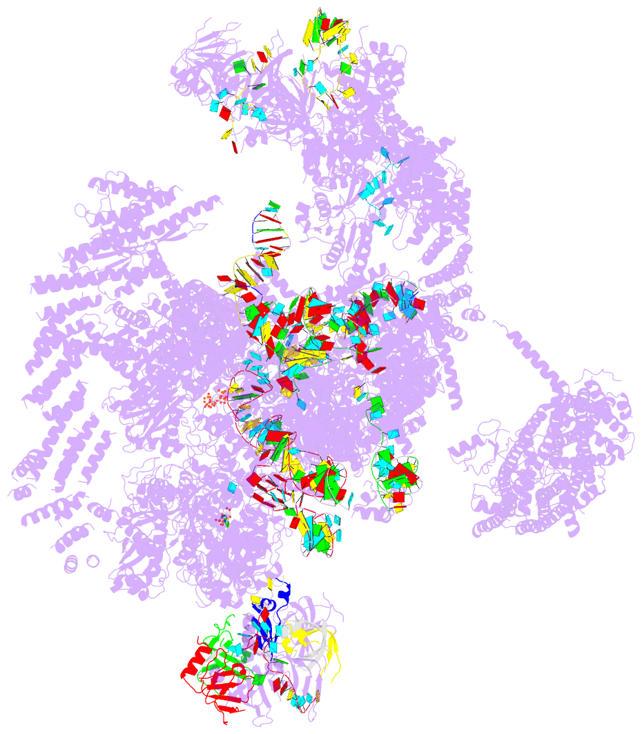
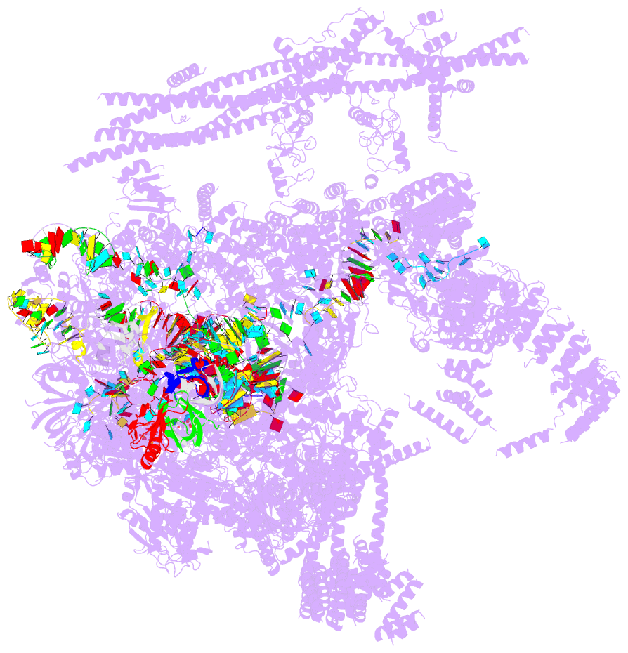
- Summary
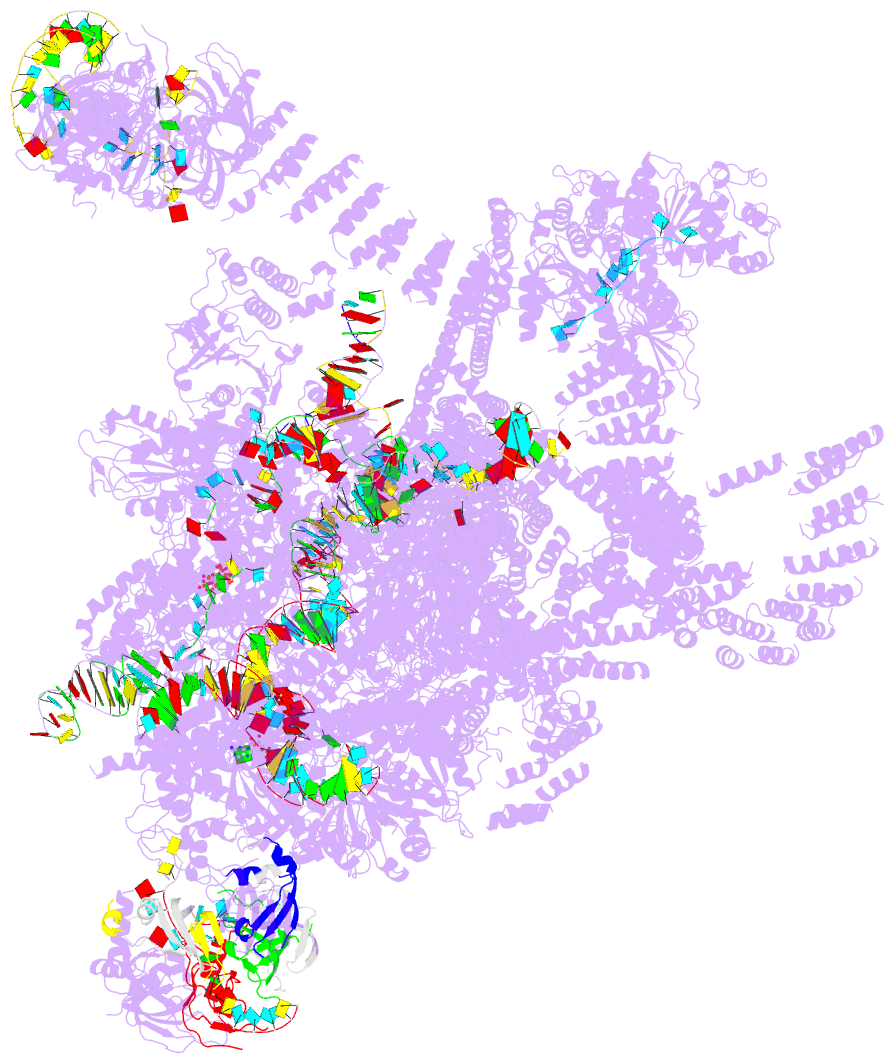
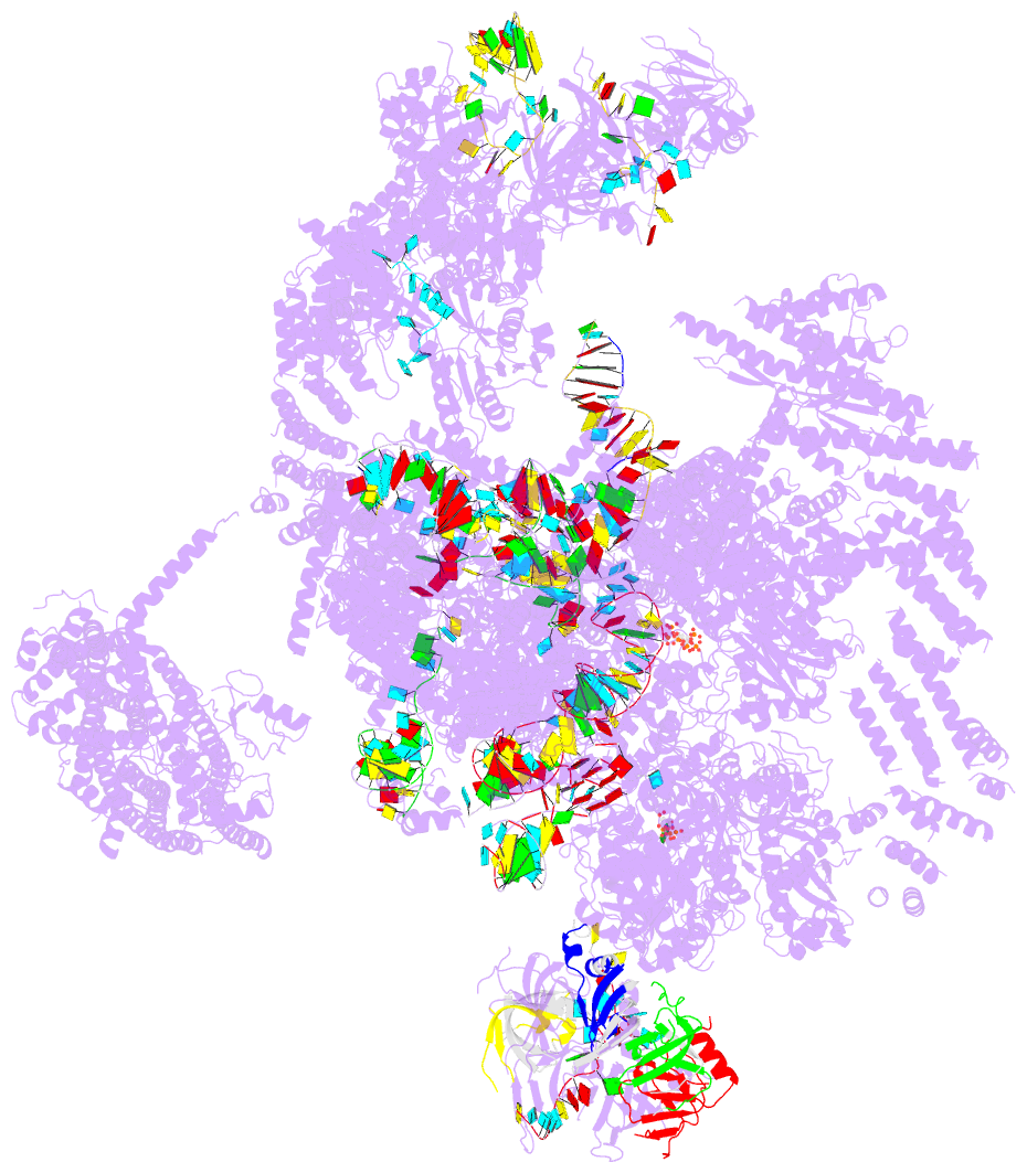
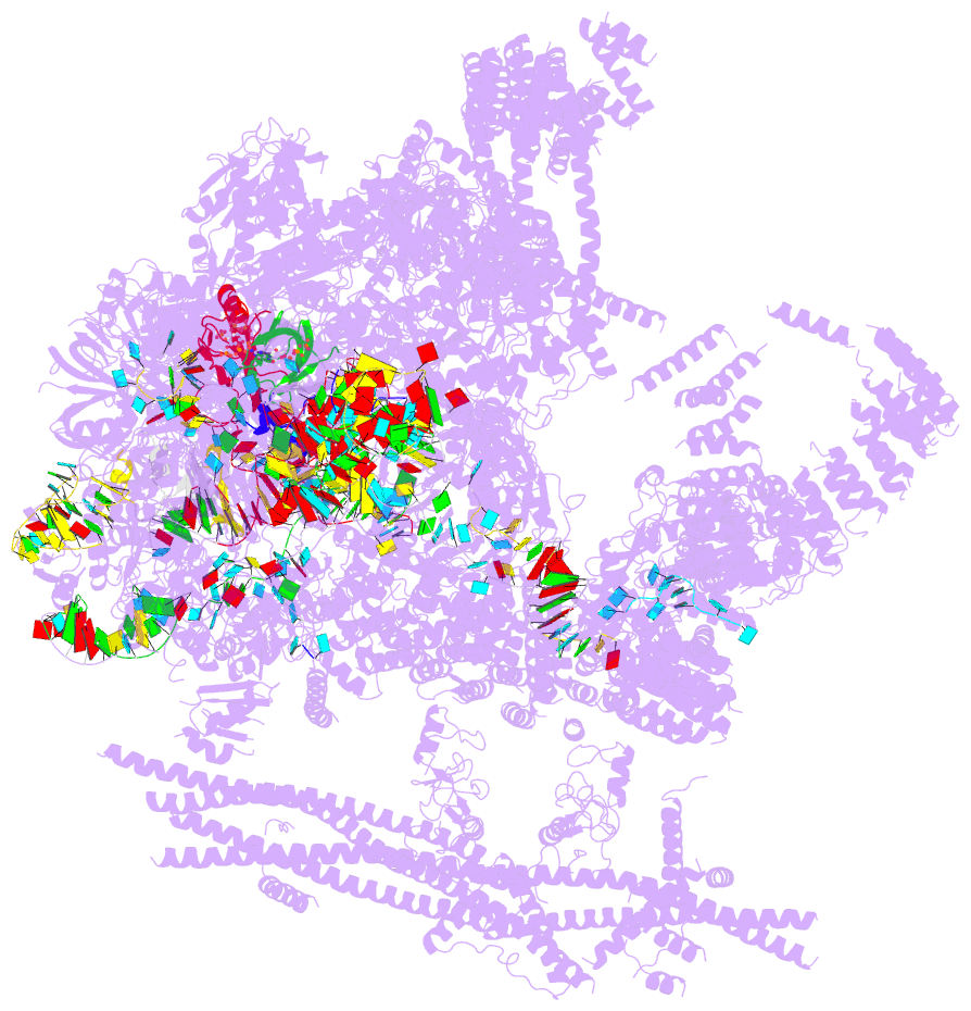
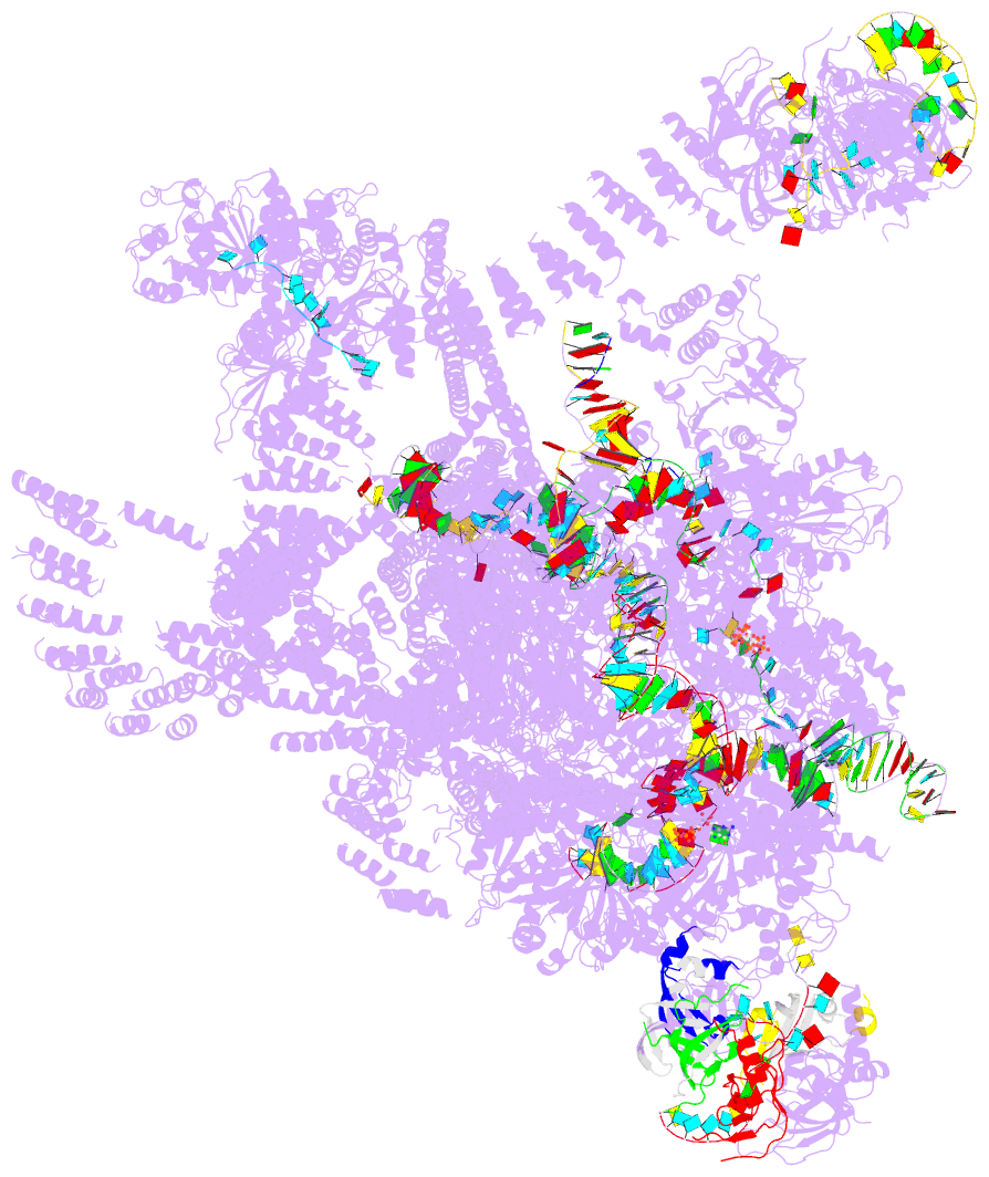
- cryo-EM structure of the intron-lariat spliceosome ready for disassembly from s.cerevisiae at 3.5 angstrom
- Reference
- Wan R, Yan C, Bai R, Lei J, Shi Y (2017): "Structure of an Intron Lariat Spliceosome from Saccharomyces cerevisiae." Cell(Cambridge,Mass.), 171, 120-132. doi: 10.1016/j.cell.2017.08.029.
- Abstract
- The disassembly of the intron lariat spliceosome (ILS) marks the end of a splicing cycle. Here we report a cryoelectron microscopy structure of the ILS complex from Saccharomyces cerevisiae at an average resolution of 3.5 Å. The intron lariat remains bound in the spliceosome whereas the ligated exon is already dissociated. The step II splicing factors Prp17 and Prp18, along with Cwc21 and Cwc22 that stabilize the 5' exon binding to loop I of U5 small nuclear RNA (snRNA), have been released from the active site assembly. The DEAH family ATPase/helicase Prp43 binds Syf1 at the periphery of the spliceosome, with its RNA-binding site close to the 3' end of U6 snRNA. The C-terminal domain of Ntr1/Spp382 associates with the GTPase Snu114, and Ntr2 is anchored to Prp8 while interacting with the superhelical domain of Ntr1. These structural features suggest a plausible mechanism for the disassembly of the ILS complex.