Summary information and primary citation
- PDB-id
- 5swd; DSSR-derived features in text and JSON formats
- Class
- RNA
- Method
- X-ray (2.5 Å)
- Summary
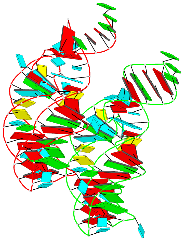
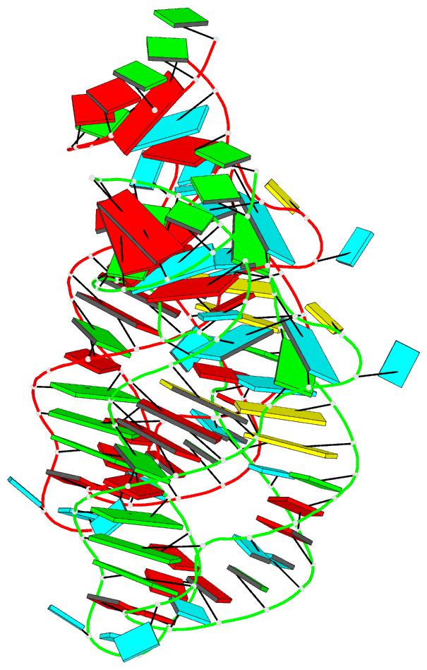
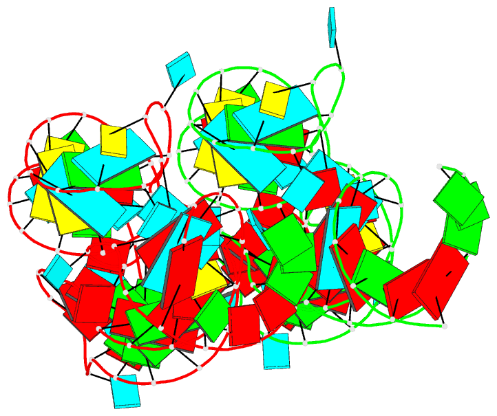
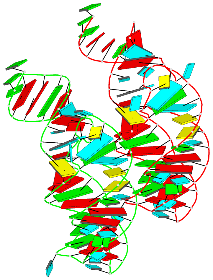
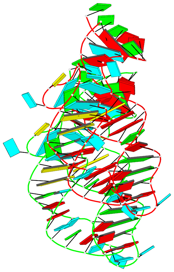
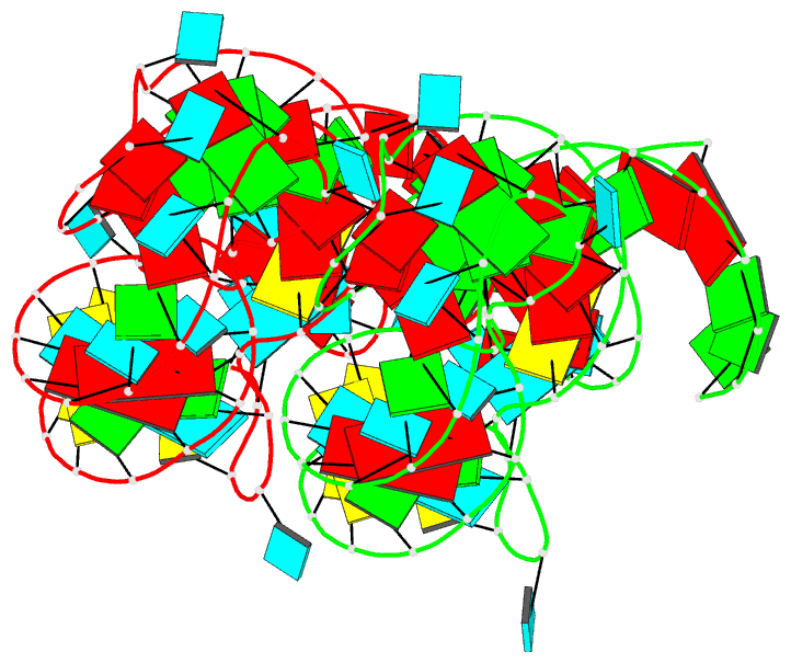
- Structure of the adenine riboswitch aptamer domain in an intermediate-bound state
- Reference
- Stagno JR, Liu Y, Bhandari YR, Conrad CE, Panja S, Swain M, Fan L, Nelson G, Li C, Wendel DR, White TA, Coe JD, Wiedorn MO, Knoska J, Oberthuer D, Tuckey RA, Yu P, Dyba M, Tarasov SG, Weierstall U, Grant TD, Schwieters CD, Zhang J, Ferre-D'Amare AR, Fromme P, Draper DE, Liang M, Hunter MS, Boutet S, Tan K, Zuo X, Ji X, Barty A, Zatsepin NA, Chapman HN, Spence JC, Woodson SA, Wang YX (2017): "Structures of riboswitch RNA reaction states by mix-and-inject XFEL serial crystallography." Nature, 541, 242-246. doi: 10.1038/nature20599.
- Abstract
- Riboswitches are RNA structural elements generally located in the 3'untranslated region (3'UTR) of mRNA. In the genetic regulation, ligand binding to the aptamer domain of a riboswitch triggers a signal to the downstream expression platform(1,2,3). A complete understanding of the structural basis for this mechanism requires the ability to study structural changes over time(4). Here we apply femtosecond X-ray free electron laser (XFEL) pulses(5,6) to obtain structural measurements from crystals so small that diffusion of a ligand can be timed to initiate a reaction prior to diffraction. We demonstrate this approach by determining four structures of the adenine riboswitch aptamer domain during the course of a reaction involving two apo, one ligand-bound intermediate, and the final bound states. These structures support a reaction mechanism model with at least four states and illustrate the structural basis for signal transmission. The two apo conformers differ significantly in the three-way junction and the P1 switch helix relative to the ligand-bound conformation. Our time-resolved crystallographic measurements with a 10-second delay captured the structure of an intermediate with changes in the binding pocket that accommodate the ligand. With a >10-minute delay, the RNA molecules were fully converted to the bound state, in which the substantial conformational changes resulted in conversion of the space group. Such drastic changes in crystallo highlight the important opportunities that micro/nanocrystals may offer in these and similar time-resolved diffraction studies. These results all together demonstrate the potential of 'mix-and-inject' time-resolved serial crystallography to study biochemically important interactions between biomacromolecules and ligands, including those involving large conformational changes.