Summary information and primary citation
- PDB-id
- 5dnb; DSSR-derived features in text and JSON formats
- Class
- DNA
- Method
- X-ray (1.4 Å)
- Summary
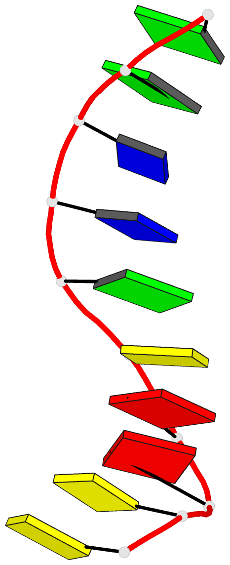
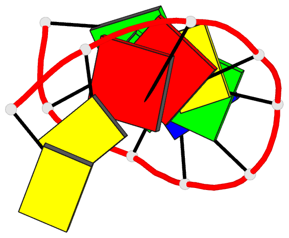
- Structure of the b-DNA decamer c-c-a-a-c-g-t-t-g-g and comparison with isomorphous decamers c-c-a-a-g-a-t-t-g-g and c-c-a-g-g-c-c-t-g-g
- Reference
- Prive GG, Yanagi K, Dickerson RE (1991): "Structure of the B-DNA decamer C-C-A-A-C-G-T-T-G-G and comparison with isomorphous decamers C-C-A-A-G-A-T-T-G-G and C-C-A-G-G-C-C-T-G-G." J.Mol.Biol., 217, 177-199. doi: 10.1016/0022-2836(91)90619-H.
- Abstract
- The crystal structure of the DNA decamer C-C-A-A-C-G-T-T-G-G has been solved to a resolution of 1.4 A, and is compared with the 1.3 A structure of C-C-A-A-G-A-T-T-G-G and the 1.6 A structure of C-C-A-G-G-C-C-T-G-G. All three decamers crystallize isomorphously in space group C2 with five base-pairs per asymmetric unit, and with decamer double helices stacked atop one another along the c axis in a manner that closely approximates a continuous B helix. This efficient stacking probably accounts for the high resolution of the crystal data. Comparison of the three decamers reveals the following. (1) Minor groove width is more variable than heretofore realized. Regions of A.T base-pairs tend to be narrower than average, although two successive A.T base-pairs alone may not be sufficient to produce narrowing. The minor groove is wider in regions where BII phosphate conformations are opposed diagonally across the groove. (2) Narrow regions of minor groove exhibit a zig-zag spine of hydration, as was first seen in C-G-C-G-A-A-T-T-C-G-C-G, whereas wide regions show two ribbons of water molecules down the walls, connecting base edge N or O with sugar O-4' atoms. Regions of intermediate groove width may accommodate neither pattern of hydration well, and may exhibit a less regular pattern of hydration. (3) Base-pair stacking is virtually identical at equivalent positions in the three decamers. The unconnected step from the top of one decamer helix to the bottom of the next helix is a normal helix step in all respects, except for the absence of connecting phosphate groups. (4) BII phosphate conformation require the unstacking of the two bases linked by the phosphate, but do not necessarily follow as an inevitable consequence of unstacking. They have an influence on minor groove width as noted in point (1) above. (5) Sugar ring pseudorotation P and main-chain torsion angle delta show an excellent correlation as given by the equation: delta = 40 degrees cos (P + 144 degrees) + 120 degrees. Although centered around C-2'-endo, the conformations in these B-DNA helices are distributed broadly from C-3'-exo to O-4'-endo, unlike the tighter clustering around C-3'-endo observed in A-DNA oligomer structures.