Summary information and primary citation
- PDB-id
- 3odc; DSSR-derived features in text and JSON formats
- Class
- DNA binding protein-DNA
- Method
- X-ray (2.8 Å)
- Summary
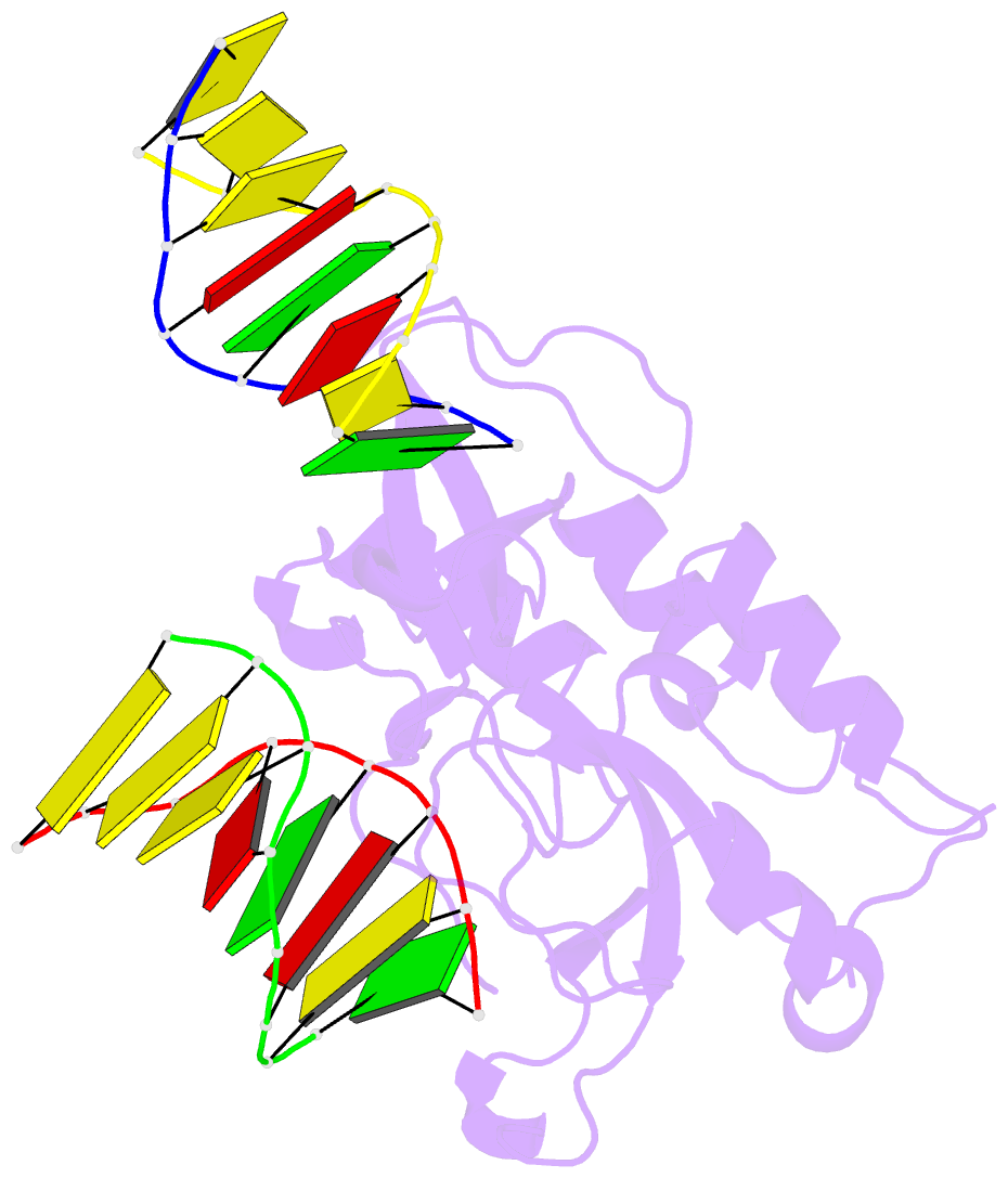
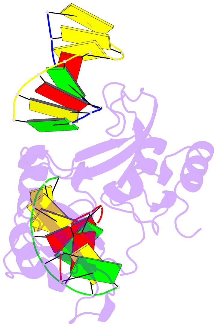
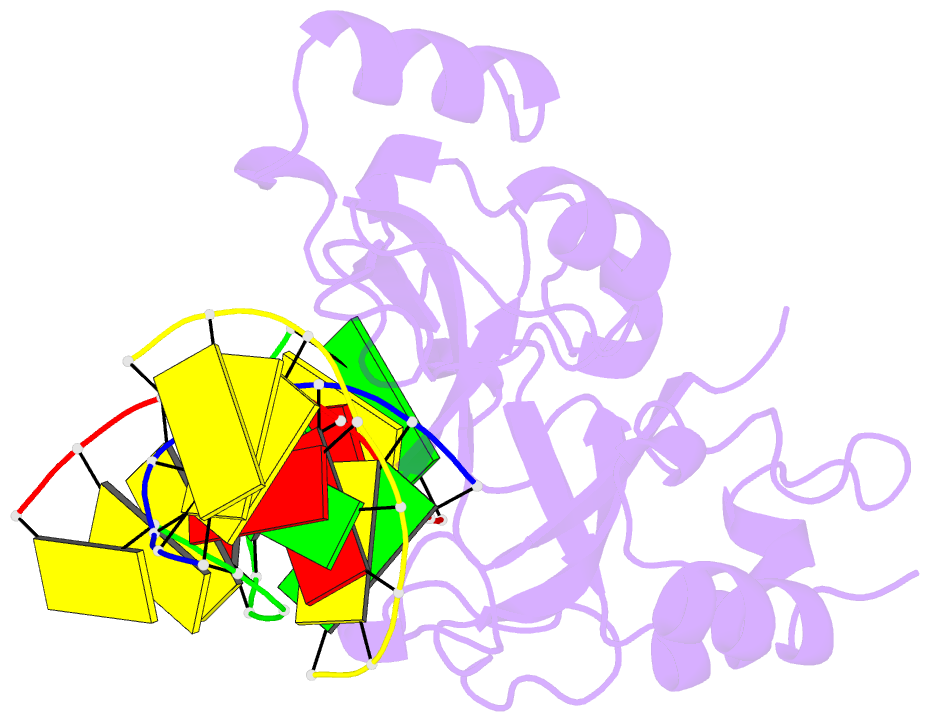
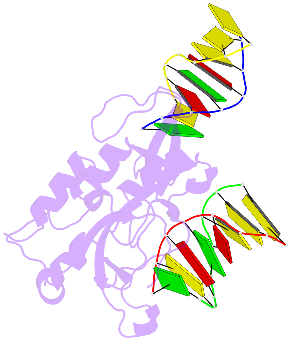
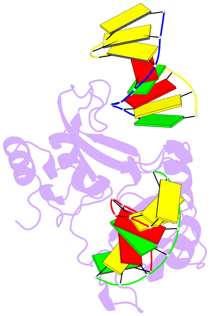
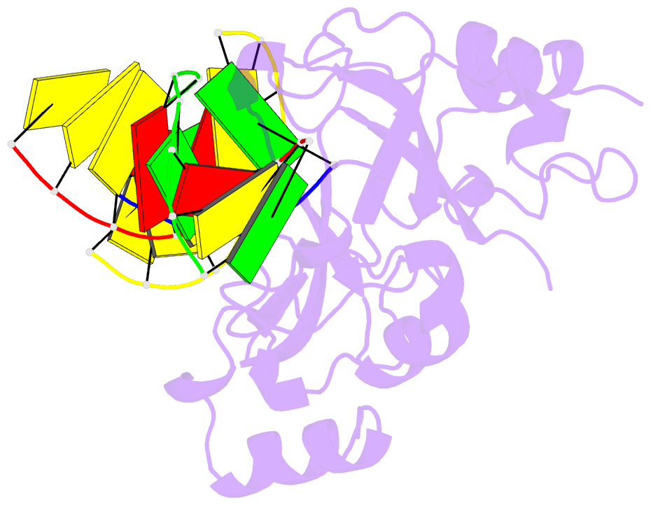
- Human parp-1 zinc finger 2 (zn2) bound to DNA
- Reference
- Langelier MF, Planck JL, Roy S, Pascal JM (2011): "Crystal Structures of Poly(ADP-ribose) Polymerase-1 (PARP-1) Zinc Fingers Bound to DNA: STRUCTURAL AND FUNCTIONAL INSIGHTS INTO DNA-DEPENDENT PARP-1 ACTIVITY." J.Biol.Chem., 286, 10690-10701. doi: 10.1074/jbc.M110.202507.
- Abstract
- Poly(ADP-ribose) polymerase-1 (PARP-1) has two homologous zinc finger domains, Zn1 and Zn2, that bind to a variety of DNA structures to stimulate poly(ADP-ribose) synthesis activity and to mediate PARP-1 interaction with chromatin. The structural basis for interaction with DNA is unknown, which limits our understanding of PARP-1 regulation and involvement in DNA repair and transcription. Here, we have determined crystal structures for the individual Zn1 and Zn2 domains in complex with a DNA double strand break, providing the first views of PARP-1 zinc fingers bound to DNA. The Zn1-DNA and Zn2-DNA structures establish a novel, bipartite mode of sequence-independent DNA interaction that engages a continuous region of the phosphodiester backbone and the hydrophobic faces of exposed nucleotide bases. Biochemical and cell biological analysis indicate that the Zn1 and Zn2 domains perform distinct functions. The Zn2 domain exhibits high binding affinity to DNA compared with the Zn1 domain. However, the Zn1 domain is essential for DNA-dependent PARP-1 activity in vitro and in vivo, whereas the Zn2 domain is not strictly required. Structural differences between the Zn1-DNA and Zn2-DNA complexes, combined with mutational and structural analysis, indicate that a specialized region of the Zn1 domain is re-configured through the hydrophobic interaction with exposed nucleotide bases to initiate PARP-1 activation.