Summary information and primary citation
- PDB-id
- 3kov; DSSR-derived features in text and JSON formats
- Class
- transcription-DNA
- Method
- X-ray (2.9 Å)
- Summary
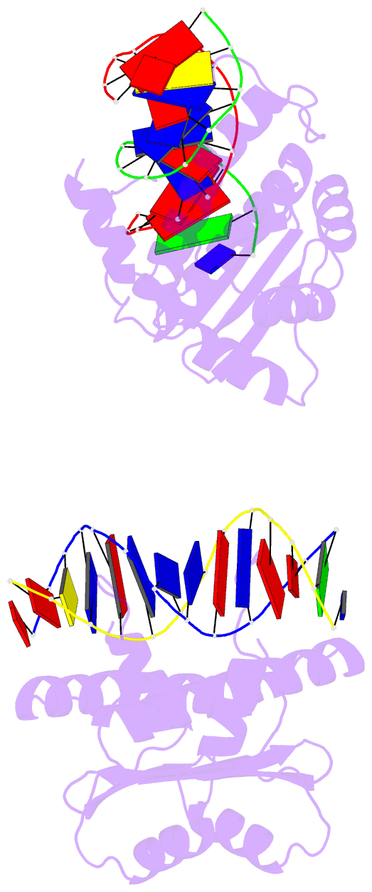
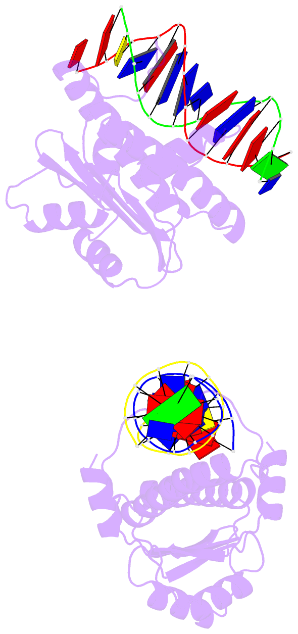
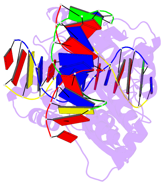
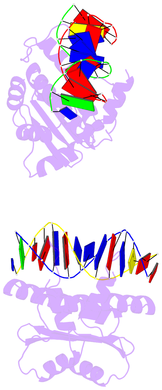
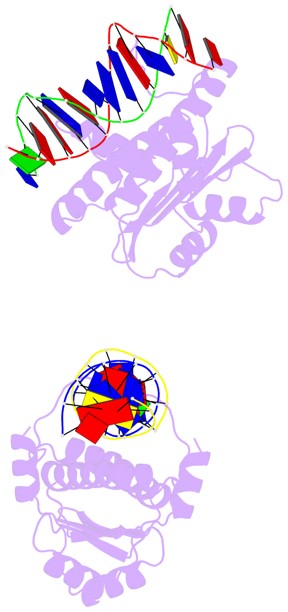
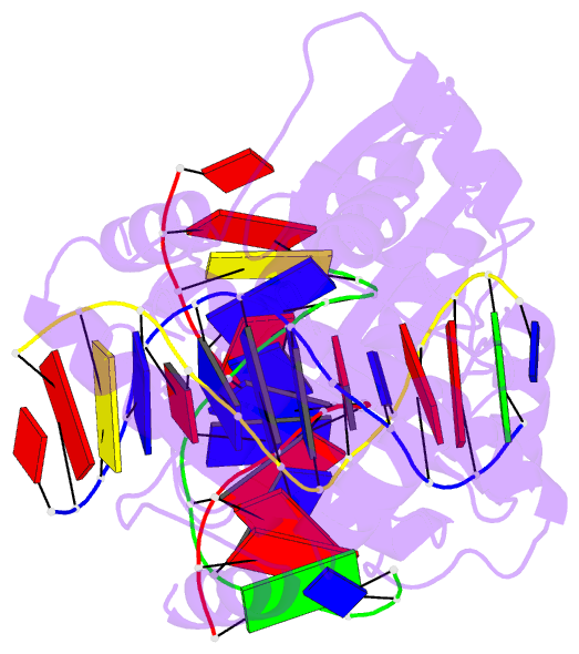
- Structure of mef2a bound to DNA reveals a completely folded mads-box-mef2 domain that recognizes DNA and recruits transcription co-factors
- Reference
- Wu Y, Dey R, Han A, Jayathilaka N, Philips M, Ye J, Chen L (2010): "Structure of the MADS-box/MEF2 Domain of MEF2A Bound to DNA and Its Implication for Myocardin Recruitment." J.Mol.Biol., 397, 520-533. doi: 10.1016/j.jmb.2010.01.067.
- Abstract
- Myocyte enhancer factor 2 (MEF2) regulates specific gene expression in diverse developmental programs and adaptive responses. MEF2 recognizes DNA and interacts with transcription cofactors through a highly conserved N-terminal domain referred to as the MADS-box/MEF2 domain. Here we present the crystal structure of the MADS-box/MEF2 domain of MEF2A bound to DNA. In contrast to previous structural studies showing that the MEF2 domain of MEF2A is partially unstructured, the present study reveals that the MEF2 domain participates with the MADS-box in both dimerization and DNA binding as a single domain. The sequence divergence at and immediately following the C-terminal end of the MEF2 domain may allow different MEF2 dimers to recognize different DNA sequences in the flanking regions. The current structure also suggests that the ligand-binding pocket previously observed in the Cabin1-MEF2B-DNA complex and the HDAC9 (histone deacetylase 9)-MEF2B-DNA complex is not induced by cofactor binding but rather preformed by intrinsic folding. However, the structure of the ligand-binding pocket does undergo subtle but significant conformational changes upon cofactor binding. On the basis of these observations, we generated a homology model of MEF2 bound to a myocardin family protein, MASTR, that acts as a potent coactivator of MEF2-dependent gene expression. The model shows excellent shape and chemical complementarity at the binding interface and is consistent with existing mutagenesis data. The apo structure presented here can also serve as a target for virtual screening and soaking studies of small molecules that can modulate the function of MEF2 as research tools and therapeutic leads.