Summary information and primary citation
- PDB-id
- 3exj; DSSR-derived features in text and JSON formats
- Class
- transcription-DNA
- Method
- X-ray (2.0 Å)
- Summary
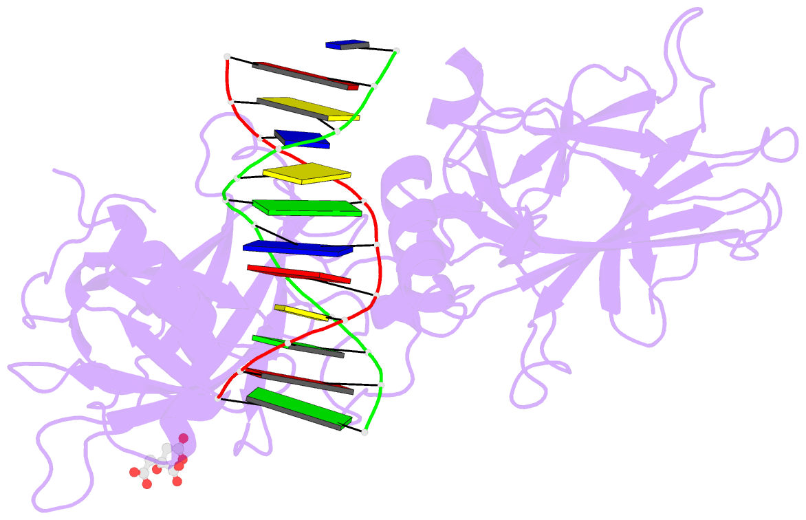
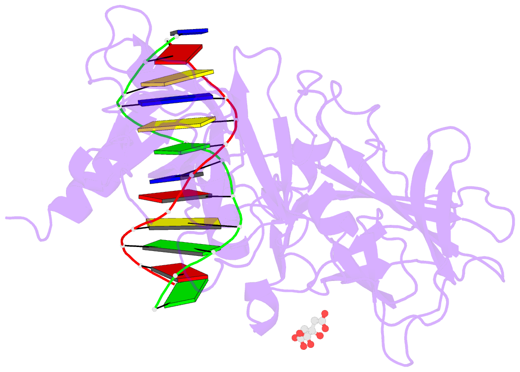
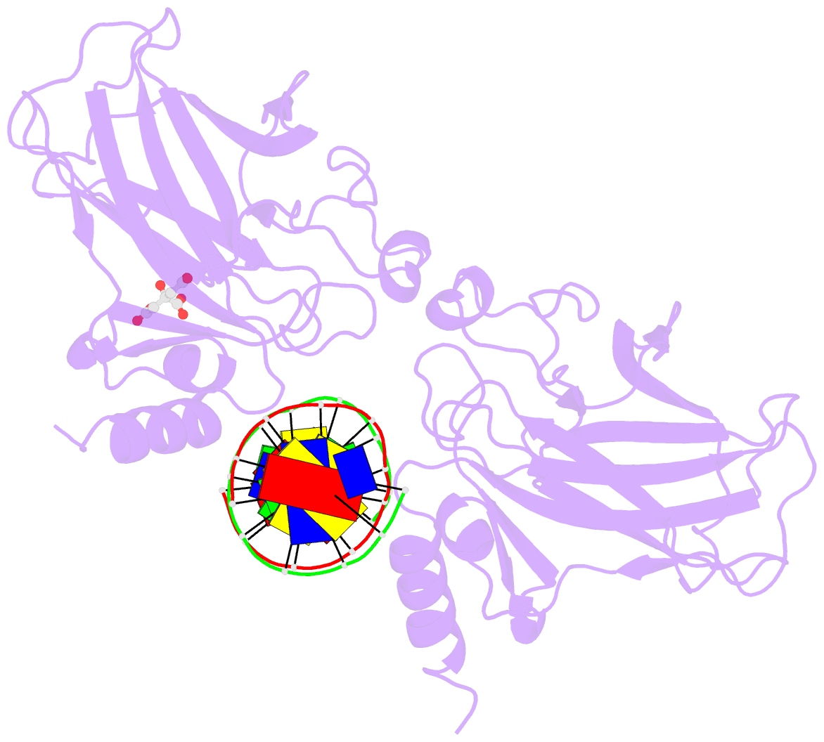
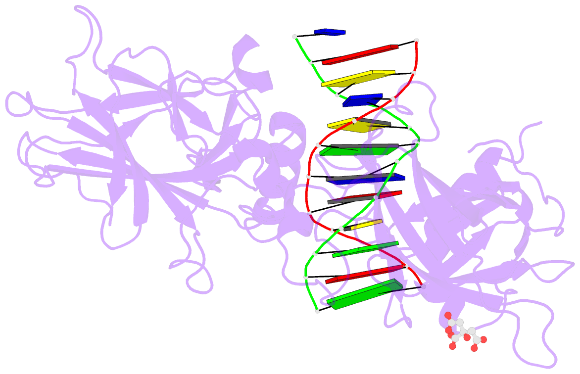
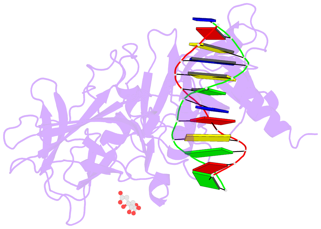
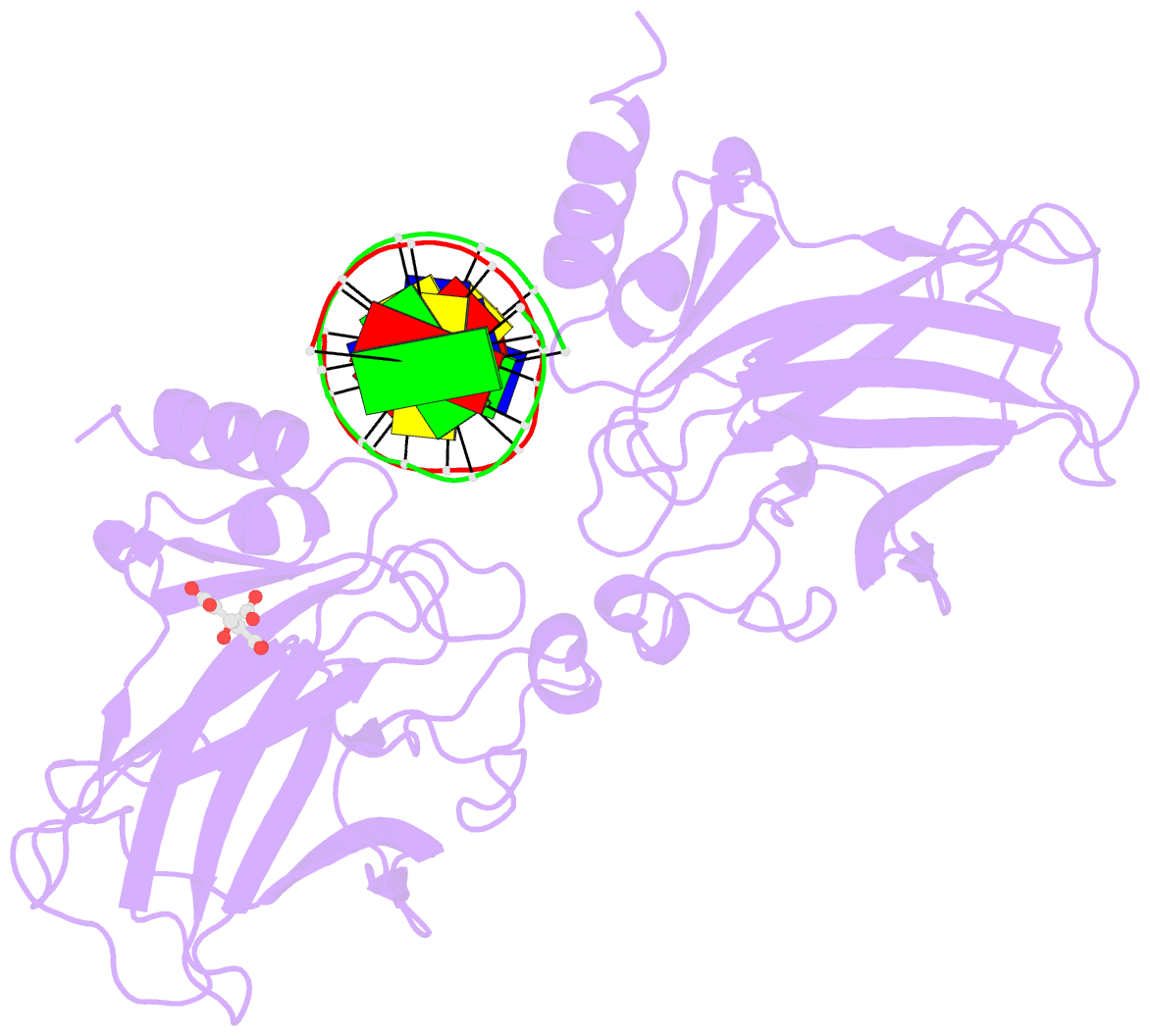
- Crystal structure of a p53 core tetramer bound to DNA
- Reference
- Malecka KA, Ho WC, Marmorstein R (2009): "Crystal structure of a p53 core tetramer bound to DNA." Oncogene, 28, 325-333. doi: 10.1038/onc.2008.400.
- Abstract
- The tumor suppressor p53 regulates downstream genes in response to many cellular stresses and is frequently mutated in human cancers. Here, we report the use of a crosslinking strategy to trap a tetrameric p53 DNA-binding domain (p53DBD) bound to DNA and the X-ray crystal structure of the protein/DNA complex. The structure reveals that two p53DBD dimers bind to B form DNA with no relative twist and that a p53 tetramer can bind to DNA without introducing significant DNA bending. The numerous dimer-dimer interactions involve several strictly conserved residues, thus suggesting a molecular basis for p53DBD-DNA binding cooperativity. Surface residue conservation of the p53DBD tetramer bound to DNA highlights possible regions of other p53 domain or p53 cofactor interactions.