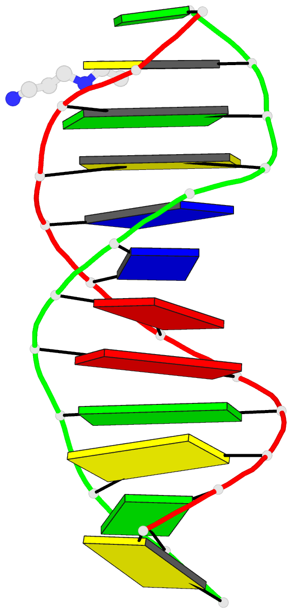
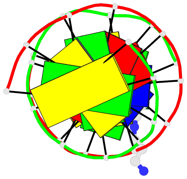
Summary information and primary citation
- PDB-id
- 355d; DSSR-derived features in text and JSON formats
- Class
- DNA
- Method
- X-ray (1.4 Å)
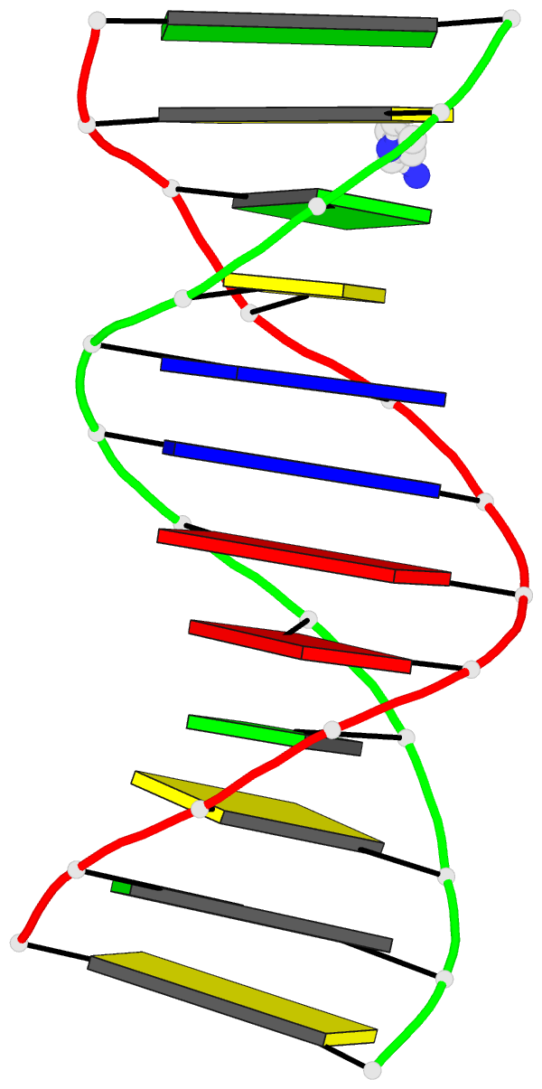
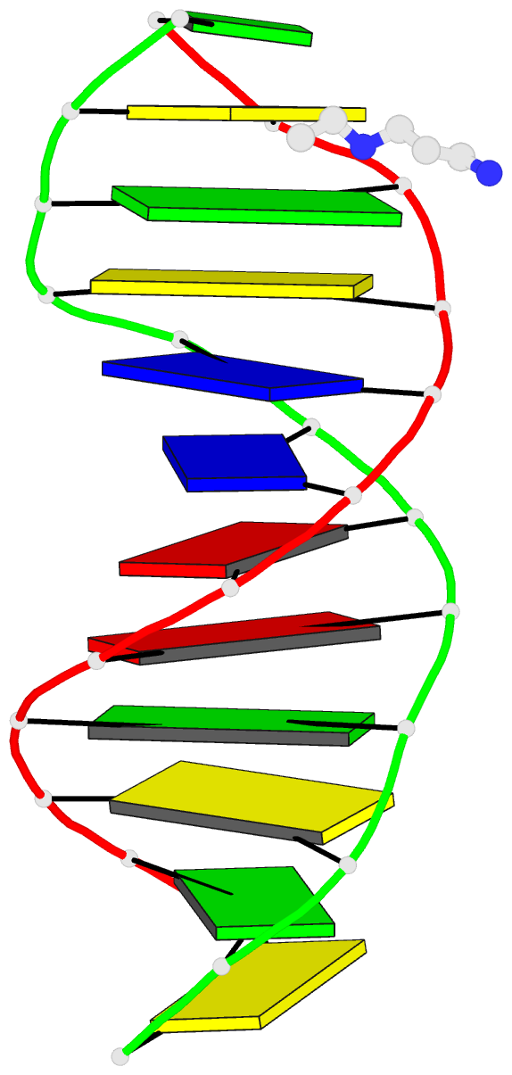
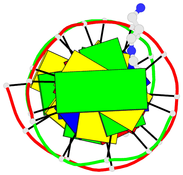
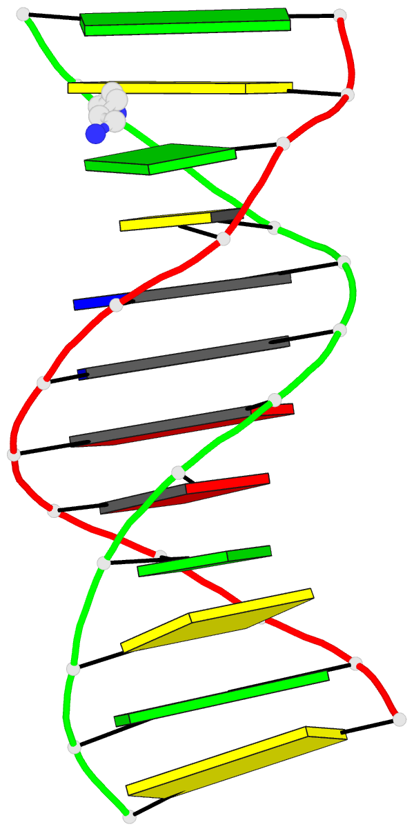
- Summary
- The b-DNA dodecamer at high resolution
- Reference
- Shui X, McFail-Isom L, Hu GG, Williams LD (1998): "The B-DNA dodecamer at high resolution reveals a spine of water on sodium." Biochemistry, 37, 8341-8355. doi: 10.1021/bi973073c.
- Abstract
- We describe a very accurate addition (called structure X here) to the B-DNA dodecamer family of X-ray structures. Our results confirm the observation of Drew and Dickerson [(1981) J. Mol. Biol. 151, 535-556] that the spine of hydration in AT tract DNA is two layers deep. However, our results suggest that the primary spine is partially occupied by sodium ions. We suggest that many sequence-dependent features of DNA conformation are mediated by site specific binding of cations. For example, preferential localization of cations, as described here within the minor groove of structure X, is probably the structural origin of AT tract bending and groove narrowing. The secondary spine, which does not interact directly with the DNA, is as geometrically regular as the primary spine, providing a model for transmission of sequence information into solvent regions. A fully hydrated magnesium ion located in the major groove of structure X appears to pull cytosine bases partially out from the helical stack, exposing pi-systems to partial positive charges of the magnesium ion and its outer sphere. A partially ordered spermine molecule is located within the major groove of structure X. Dodecamer structures are derived from crystals of [d(CGCGAATTCGCG)]2 in space group P212121 (a = 25 A, b = 40 A, and c = 66 A). On average, those crystals diffracted to around 2.5 A resolution with 2500 unique reflections. Structure X, with the same space group, DNA sequence, and crystal form as the "Dickerson dodecamer", is refined against a complete, low-temperature, 1.4 A resolution data set, with over 11000 reflections. Structure X appears to be conformationally more ordered than previous structures, suggesting that at least a portion of the conformational heterogeneity previously attributed to DNA sequence in fact arises from experimental error.