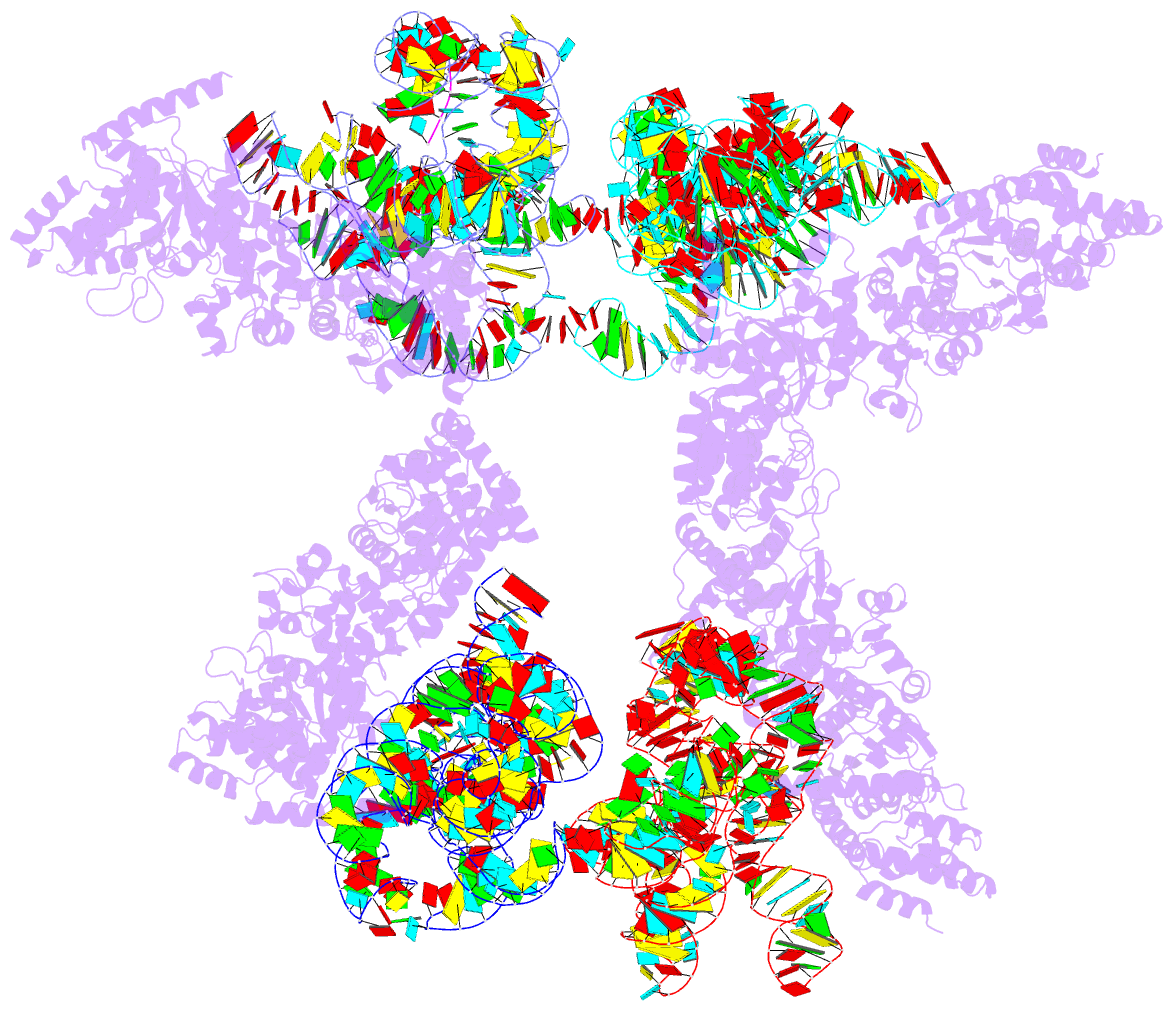
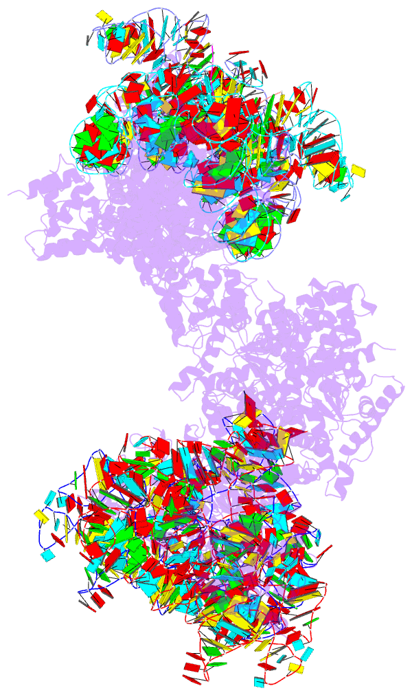
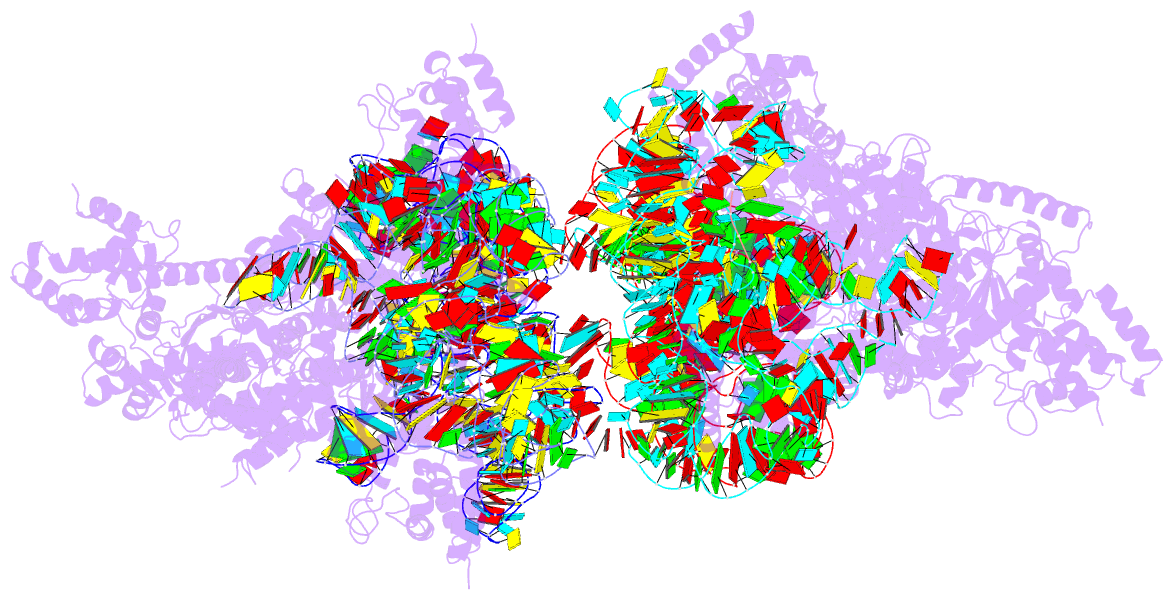
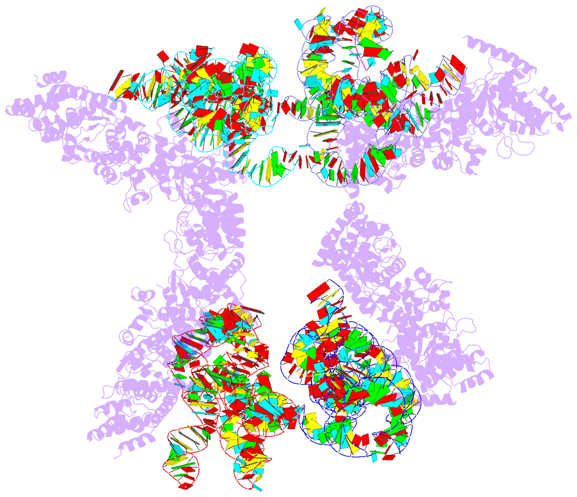
Summary information and primary citation
- PDB-id
- 2rkj; DSSR-derived features in text and JSON formats
- Class
- ligase-RNA
- Method
- X-ray (4.5 Å)
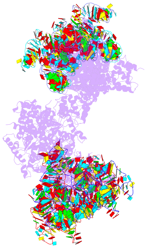
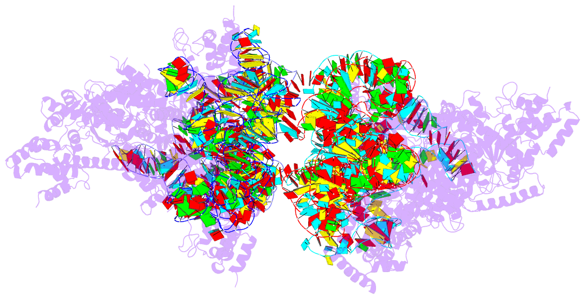
- Summary
- Cocrystal structure of a tyrosyl-trna synthetase splicing factor with a group i intron RNA
- Reference
- Paukstelis PJ, Chen JH, Chase E, Lambowitz AM, Golden BL (2008): "Structure of a tyrosyl-tRNA synthetase splicing factor bound to a group I intron RNA." Nature, 451, 94-97. doi: 10.1038/nature06413.
- Abstract
- The 'RNA world' hypothesis holds that during evolution the structural and enzymatic functions initially served by RNA were assumed by proteins, leading to the latter's domination of biological catalysis. This progression can still be seen in modern biology, where ribozymes, such as the ribosome and RNase P, have evolved into protein-dependent RNA catalysts ('RNPzymes'). Similarly, group I introns use RNA-catalysed splicing reactions, but many function as RNPzymes bound to proteins that stabilize their catalytically active RNA structure. One such protein, the Neurospora crassa mitochondrial tyrosyl-tRNA synthetase (TyrRS; CYT-18), is bifunctional and both aminoacylates mitochondrial tRNA(Tyr) and promotes the splicing of mitochondrial group I introns. Here we determine a 4.5-A co-crystal structure of the Twort orf142-I2 group I intron ribozyme bound to splicing-active, carboxy-terminally truncated CYT-18. The structure shows that the group I intron binds across the two subunits of the homodimeric protein with a newly evolved RNA-binding surface distinct from that which binds tRNA(Tyr). This RNA binding surface provides an extended scaffold for the phosphodiester backbone of the conserved catalytic core of the intron RNA, allowing the protein to promote the splicing of a wide variety of group I introns. The group I intron-binding surface includes three small insertions and additional structural adaptations relative to non-splicing bacterial TyrRSs, indicating a multistep adaptation for splicing function. The co-crystal structure provides insight into how CYT-18 promotes group I intron splicing, how it evolved to have this function, and how proteins could have incrementally replaced RNA structures during the transition from an RNA world to an RNP world.