Summary information and primary citation
- PDB-id
- 2meq; DSSR-derived features in text and JSON formats
- Class
- RNA
- Method
- NMR
- Summary
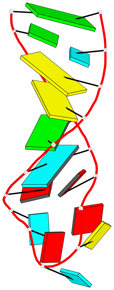
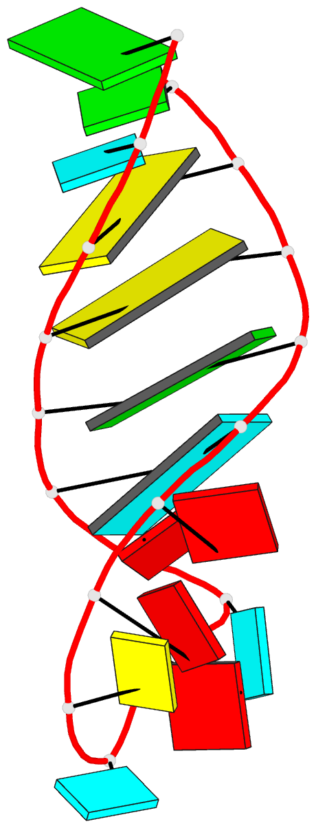
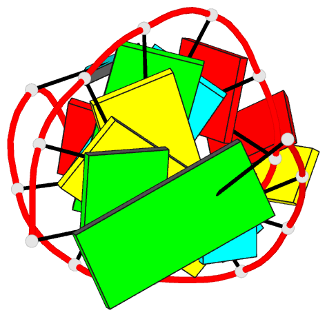
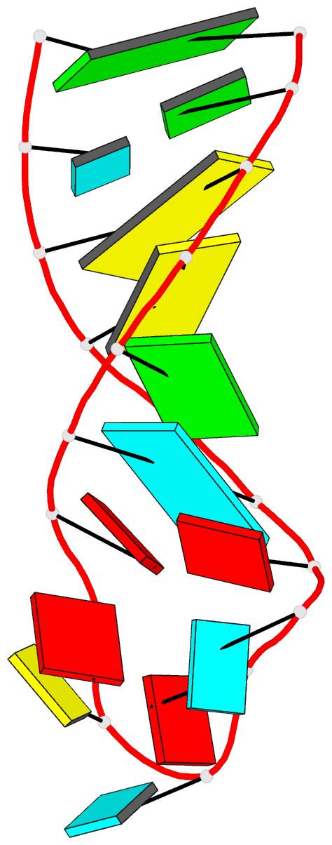
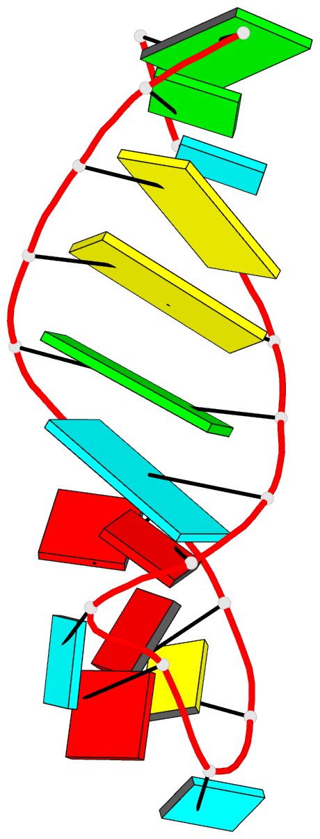
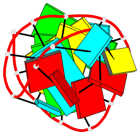
- Structure of helix 69 from escherichia coli 23s ribosomal RNA
- Reference
- Jiang J, Aduri R, Chow CS, Santalucia J (2014): "Structure modulation of helix 69 from Escherichia coli 23S ribosomal RNA by pseudouridylations." Nucleic Acids Res., 42, 3971-3981. doi: 10.1093/nar/gkt1329.
- Abstract
- Helix 69 (H69) is a 19-nt stem-loop region from the large subunit ribosomal RNA. Three pseudouridine (Ψ) modifications clustered in H69 are conserved across phylogeny and known to affect ribosome function. To explore the effects of Ψ on the conformations of Escherichia coli H69 in solution, nuclear magnetic resonance spectroscopy was used to reveal the structural differences between H69 with (ΨΨΨ) and without (UUU) Ψ modifications. Comparison of the two structures shows that H69 ΨΨΨ has the following unique features: (i) the loop region is closed by a Watson-Crick base pair between Ψ1911 and A1919, which is potentially reinforced by interactions involving Ψ1911N1H and (ii) Ψ modifications at loop residues 1915 and 1917 promote base stacking from Ψ1915 to A1918. In contrast, the H69 UUU loop region, which lacks Ψ modifications, is less organized. Structure modulation by Ψ leads to alteration in conformational behavior of the 5' half of the H69 loop region, observed as broadening of C1914 non-exchangeable base proton resonances in the H69 ΨΨΨ nuclear magnetic resonance spectra, and plays an important biological role in establishing the ribosomal intersubunit bridge B2a and mediating translational fidelity.