Summary information and primary citation
- PDB-id
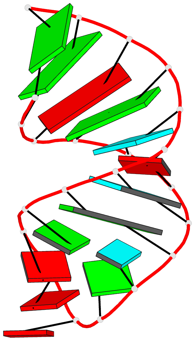
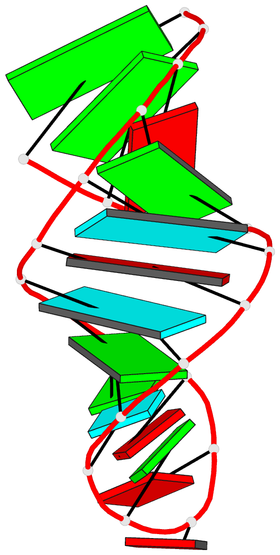
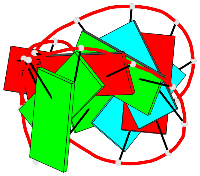
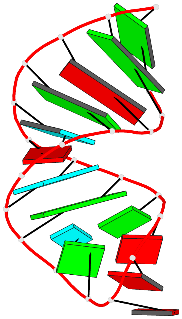
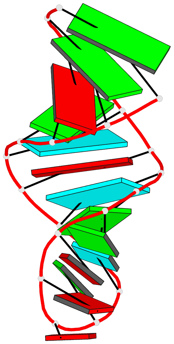
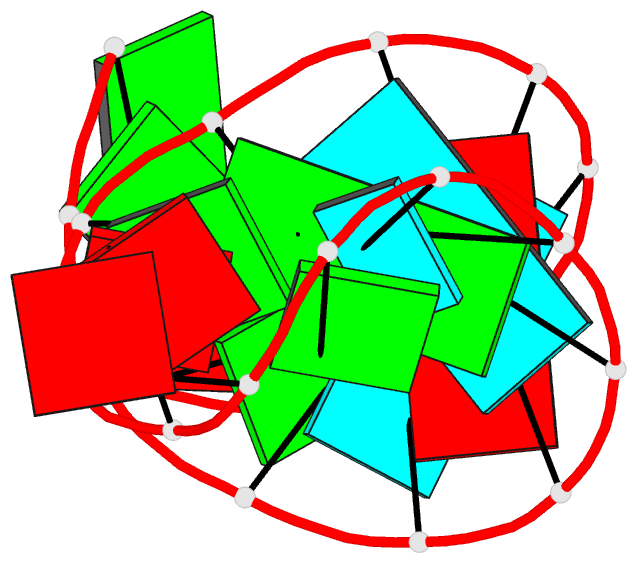
- 2k66; DSSR-derived features in text and JSON formats
- Class
- RNA
- Method
- NMR
- Summary
- NMR solution structure of the d3'-stem closed by a gaaa tetraloop of the group ii intron sc.ai5(gamma)
- Reference
- Kruschel D, Skilandat M, Sigel RK (2014): "NMR structure of the 5' splice site in the group IIB intron Sc.ai5 gamma--conformational requirements for exon-intron recognition." Rna, 20, 295-307. doi: 10.1261/rna.041137.113.
- Abstract
- A crucial step of the self-splicing reaction of group II intron ribozymes is the recognition of the 5' exon by the intron. This recognition is achieved by two regions in domain 1 of the intron, the exon-binding sites EBS1 and EBS2 forming base pairs with the intron-binding sites IBS1 and IBS2 located at the end of the 5' exon. The complementarity of the EBS1•IBS1 contact is most important for ensuring site-specific cleavage of the phosphodiester bond between the 5' exon and the intron. Here, we present the NMR solution structures of the d3' hairpin including EBS1 free in solution and bound to the IBS1 7-mer. In the unbound state, EBS1 is part of a flexible 11-nucleotide (nt) loop. Binding of IBS1 restructures and freezes the entire loop region. Mg(2+) ions are bound near the termini of the EBS1•IBS1 helix, stabilizing the interaction. Formation of the 7-bp EBS1•IBS1 helix within a loop of only 11 nt forces the loop backbone to form a sharp turn opposite of the splice site, thereby presenting the scissile phosphate in a position that is structurally unique.