Summary information and primary citation
- PDB-id
- 2hax; DSSR-derived features in text and JSON formats
- Class
- gene regulation-DNA
- Method
- X-ray (1.29 Å)
- Summary
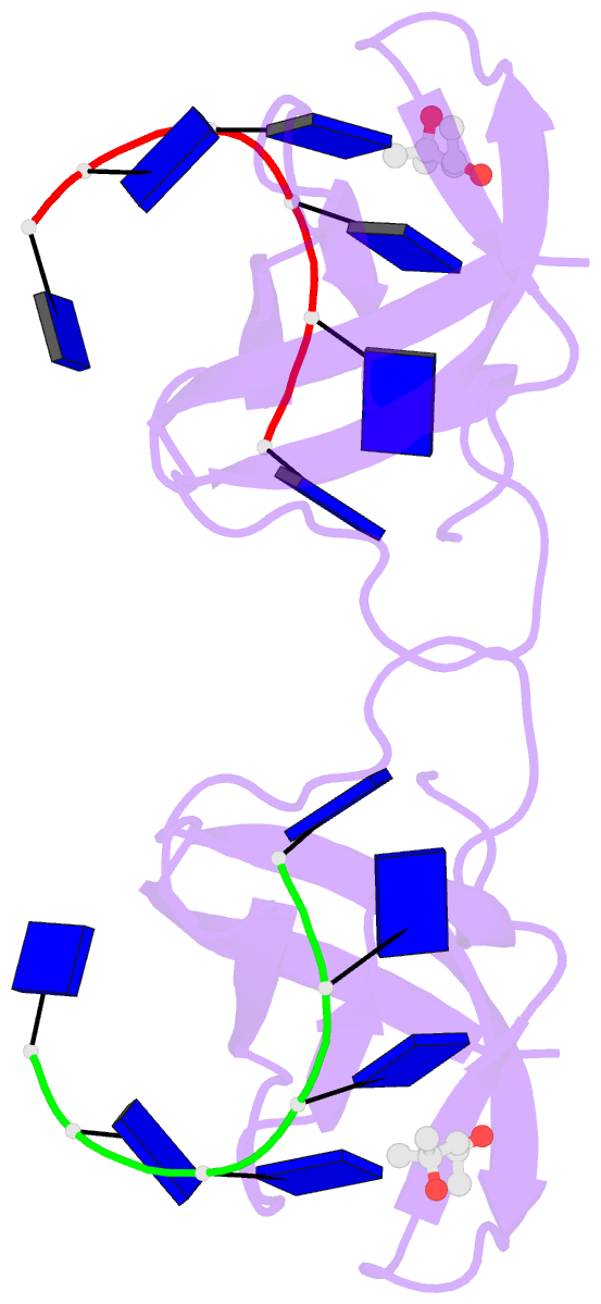
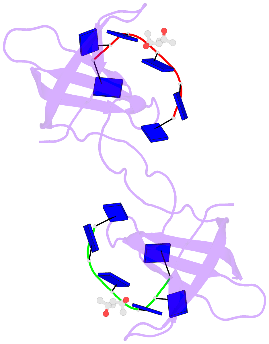
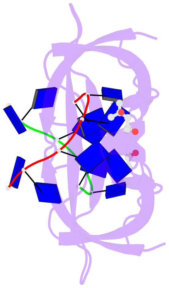
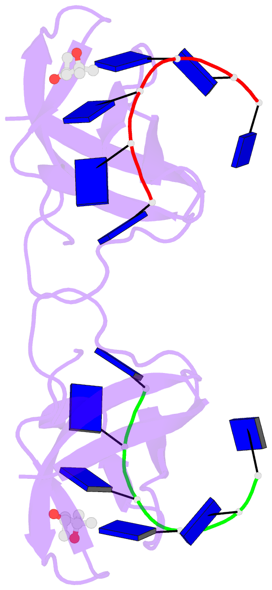
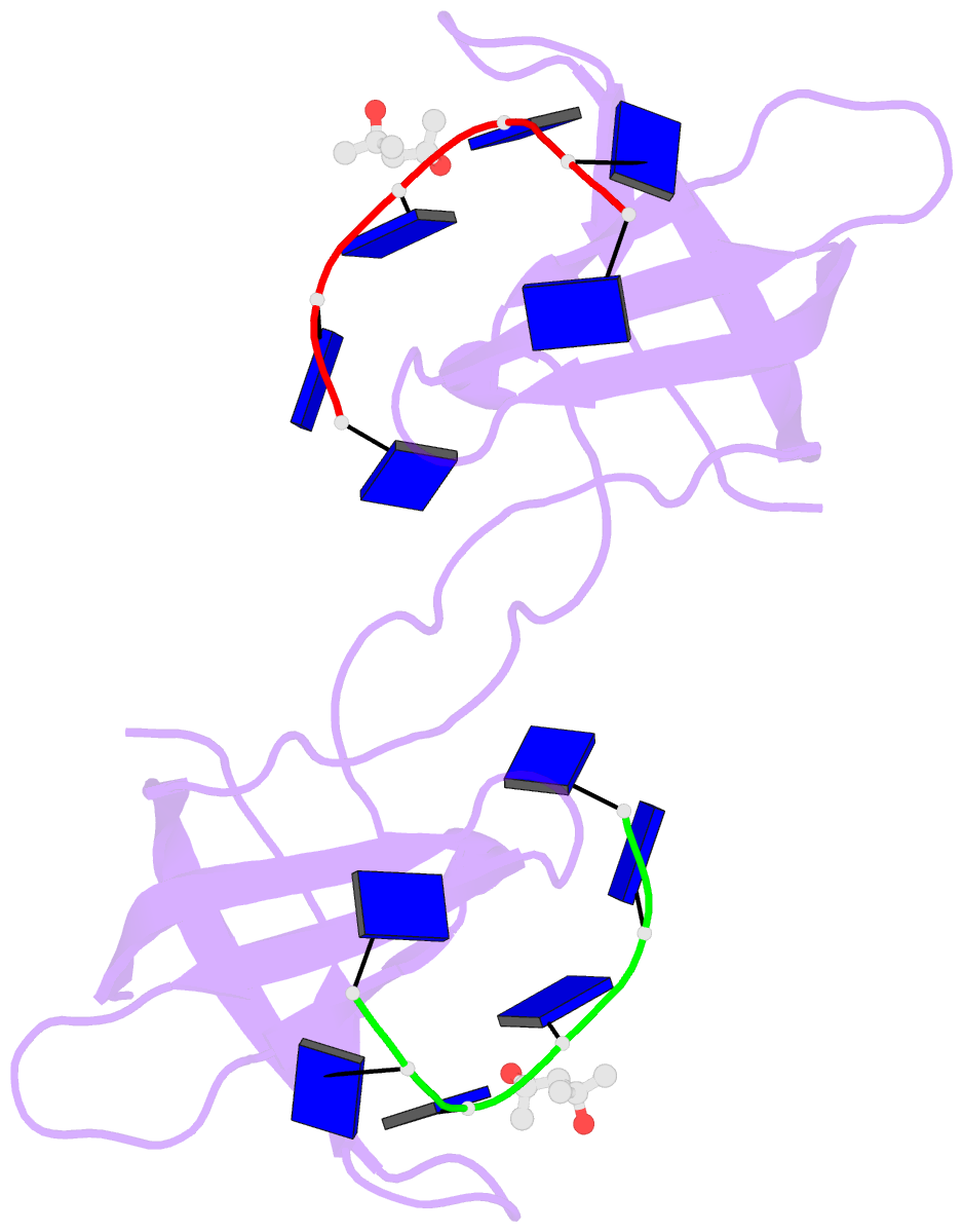
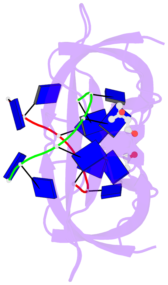
- Crystal structure of bacillus caldolyticus cold shock protein in complex with hexathymidine
- Reference
- Max KE, Zeeb M, Bienert R, Balbach J, Heinemann U (2007): "Common mode of DNA binding to cold shock domains. Crystal structure of hexathymidine bound to the domain-swapped form of a major cold shock protein from Bacillus caldolyticus." Febs J., 274, 1265-1279. doi: 10.1111/j.1742-4658.2007.05672.x.
- Abstract
- Bacterial cold shock proteins (CSPs) regulate cellular adaptation to cold stress. Functions ascribed to CSP include roles as RNA chaperones and in transcription antitermination. We present the crystal structure of the Bacillus caldolyticus CSP (Bc-Csp) in complex with hexathymidine (dT(6)) at a resolution of 1.29 A. Bound to dT(6), crystalline Bc-Csp forms a domain-swapped dimer in which beta strands 1-3 associate with strands 4 and 5 from the other subunit to form a closed beta barrel and vice versa. The globular units of dimeric Bc-Csp closely resemble the well-known structure of monomeric CSP. Structural reorganization from the monomer to the domain-swapped dimer involves a strictly localized change in the peptide bond linking Glu36 and Gly37 of Bc-Csp. Similar structural reorganizations have not been found in any other CSP or oligonucleotide/oligosaccharide-binding fold structures. Each dT(6) ligand is bound to one globular unit of Bc-Csp via an amphipathic protein surface. Individual binding subsites interact with the DNA bases through stacking and hydrogen bonding. The sugar-phosphate backbone remains solvent exposed. Based on crystallographic and biochemical studies of deoxyoligonucleotide binding to CSP, we suggest a common mode of binding of single-stranded heptanucleotide motifs to proteins containing cold shock domains, including the eukaryotic Y-box factors.