Summary information and primary citation
- PDB-id
-
2grb;
SNAP-derived features in text and
JSON formats
- Class
- RNA
- Method
- X-ray (1.4 Å)
- Summary
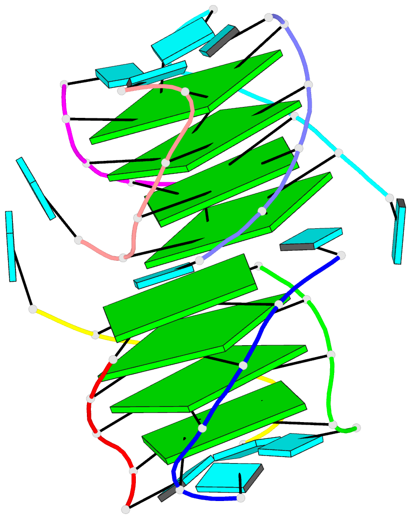
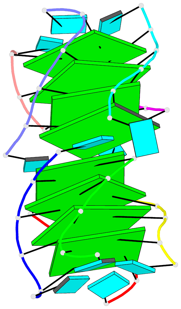
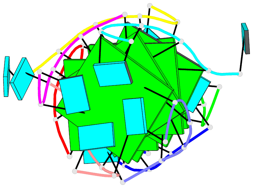
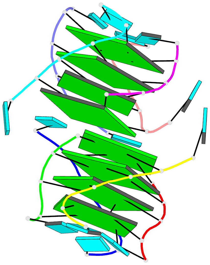
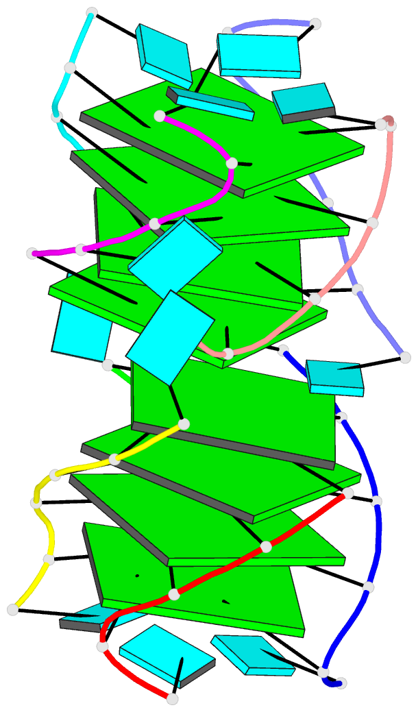
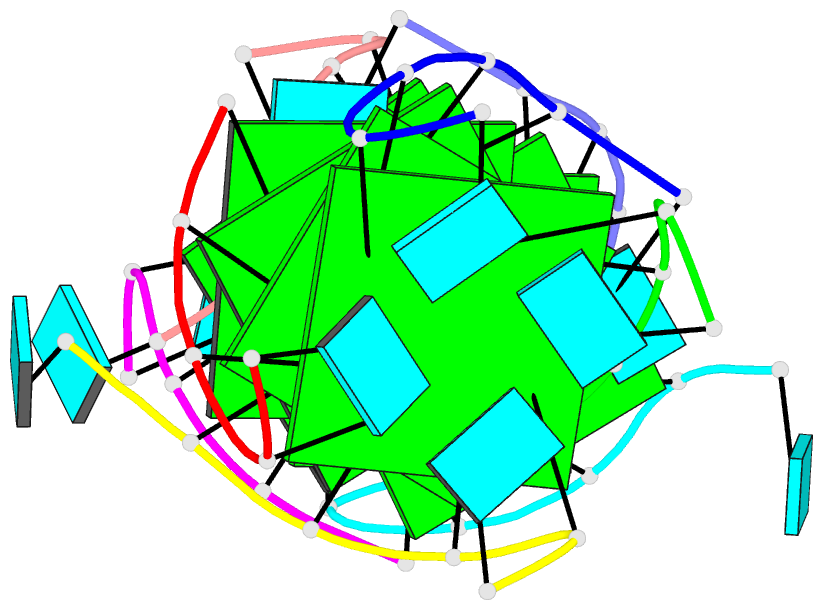
- Crystal structure of an RNA quadruplex containing
inosine-tetrad
- Reference
-
Pan B, Shi K, Sundaralingam M (2006): "Crystal
structure of an RNA quadruplex containing inosine tetrad:
implications for the roles of NH2 group in purine
tetrads." J.Mol.Biol., 363,
451-459. doi: 10.1016/j.jmb.2006.08.022.
- Abstract
- Polyinosinic acid has been known to adopt the
four-stranded helical structure but its basic unit, inosine
tetrad (I tetrad), has not been determined at the atomic
level. Here we report the crystal structure of an RNA
quadruplex containing an I tetrad at 1.4 A resolution. The
I tetrad has one cyclic hydrogen bond N1...O6 with the bond
length of 2.7 A. A water bridge is observed in the minor
groove side of the base tetrad. Even though it is
sandwiched by guanine tetrads (G tetrads), the I tetrad is
buckled towards the 3' side of the tetrad plane, which
results from the different interaction strength with K ions
on two sides of the tetrad plane. Comparison with both G
tetrad and adenine tetrad indicates that lack of NH2 in the
C2 position makes the I tetrad prone to buckle for
interactions with ligands. Two U*(G-G-G-G) base pentads are
observed at the junction of the 5' termini of two
quadruplexes. The uridine residue in the base pentad is
engaged in two hydrogen bonding interactions
(N2(G)-H...O2(U) and O2'(G)-H...O4(U)) and a water-mediated
interaction (N3(G) and N3(U)) with the G tetrad. We also
discuss the roles of amino group in purine tetrads and the
inter-quadruplex interactions in RNA molecules. These
quadruplexes may interact with each other by stacking,
groove binding and intercalation.