Summary information and primary citation
- PDB-id
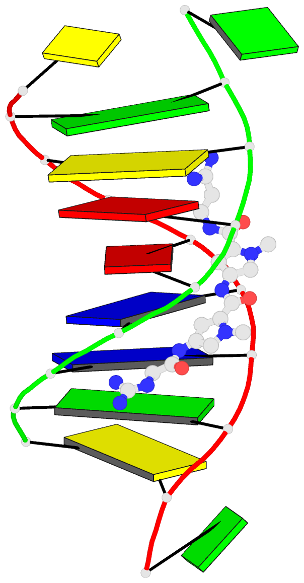
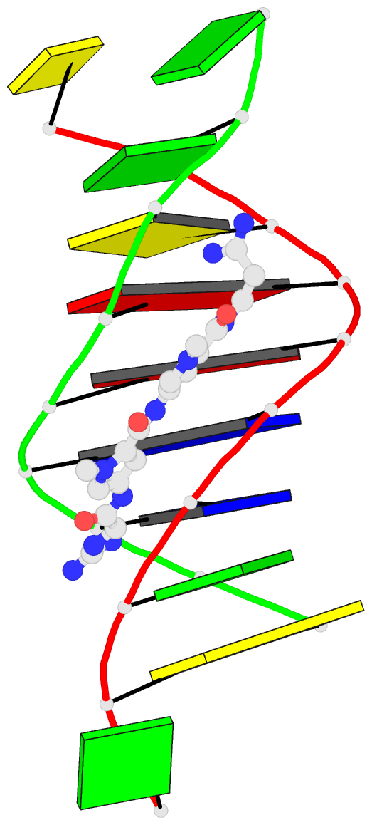
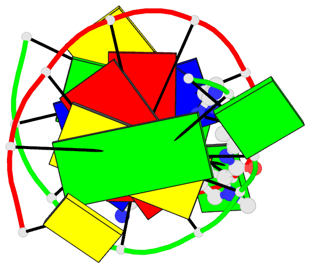
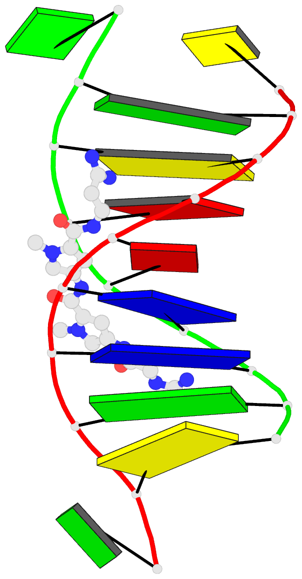
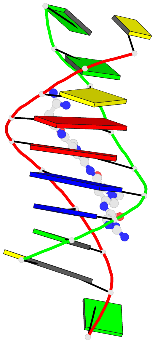
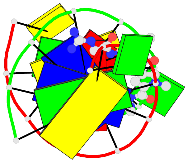
- 261d; DSSR-derived features in text and JSON formats
- Class
- DNA
- Method
- X-ray (2.4 Å)
- Summary
- Crystal structure of the DNA decamer d(cgcaattgcg) complexed with the minor groove binding drug netropsin
- Reference
- Nunn CM, Garman E, Neidle S (1997): "Crystal structure of the DNA decamer d(CGCAATTGCG) complexed with the minor groove binding drug netropsin." Biochemistry, 36, 4792-4799. doi: 10.1021/bi9628228.
- Abstract
- The crystal structure of netropsin bound to the decamer d(CGCAATTGCG) has been determined at 2.4 A resolution. This is the first example of a crystal structure of netropsin bound to decamer DNA. The central eight bases of each DNA single-strand base pair with a self-complementary strand to form an octamer B-DNA duplex. These duplexes lie end to end within the unit cell. The terminal 5'-C and G-3' bases are unpaired and interact with the neighboring duplexes via interactions within both the major and minor groove to form base triplet interactions of the type C(+)-G x C and G*(G x C), respectively. The triplet interaction of the type C(+)-G x C is known to exist within triplex DNA with the C+ base oriented parallel with the Watson-Crick guanine base to which it hydrogen bonds. The netropsin molecule lies within the minor groove of the octamer duplex and assumes a class I type position, with bifurcated hydrogen-bonding interactions from the amide groups of the netropsin to the A x T base pairs of the minor groove. The netropsin molecule fits within a five base pair long minor groove site by bending of the flexible amidinium group at one end of the drug.