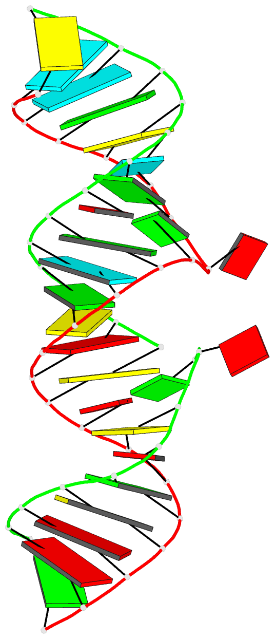
Summary information and primary citation
- PDB-id
- 1wvd; DSSR-derived features in text and JSON formats
- Class
- RNA
- Method
- X-ray (2.93 Å)
- Summary
- Hiv-1 dis(mal) duplex cocl2-soaked
- Reference
- Ennifar E, Walter P, Dumas P (2003): "A crystallographic study of the binding of 13 metal ions to two related RNA duplexes." Nucleic Acids Res., 31, 2671-2682. doi: 10.1093/nar/gkg350.
- Abstract
- Metal ions, and magnesium in particular, are known to be involved in RNA folding by stabilizing secondary and tertiary structures, and, as cofactors, in RNA enzymatic activity. We have conducted a systematic crystallographic analysis of cation binding to the duplex form of the HIV-1 RNA dimerization initiation site for the subtype-A and -B natural sequences. Eleven ions (K+, Pb2+, Mn2+, Ba2+, Ca2+, Cd2+, Sr2+, Zn2+, Co2+, Au3+ and Pt4+) and two hexammines [Co (NH3)6]3+ and [Ru (NH3)6]3+ were found to bind to the DIS duplex structure. Although the two sequences are very similar, strong differences were found in their cation binding properties. Divalent cations bind almost exclusively, as Mg2+, at 'Hoogsteen' sites of guanine residues, with a cation-dependent affinity for each site. Notably, a given cation can have very different affinities for a priori equivalent sites within the same molecule. Surprisingly, none of the two hexammines used were able to efficiently replace hexahydrated magnesium. Instead, [Co (NH3)4]3+ was seen bound by inner-sphere coordination to the RNA. This raises some questions about the practical use of [Co (NH3)6]3+ as a [Mg (H2O)6]2+ mimetic. Also very unexpected was the binding of the small Au3+ cation exactly between the Watson-Crick sites of a G-C base pair after an obligatory deprotonation of N1 of the guanine base. This extensive study of metal ion binding using X-ray crystallography significantly enriches our knowledge on the binding of middleweight or heavy metal ions to RNA, particularly compared with magnesium.


