Summary information and primary citation
- PDB-id
- 1vtf; DSSR-derived features in text and JSON formats
- Class
- DNA
- Method
- X-ray (2.0 Å)
- Summary
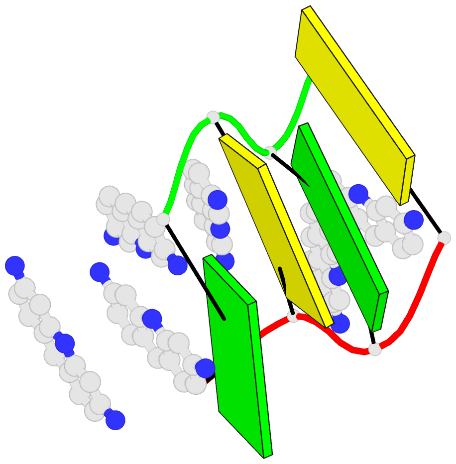
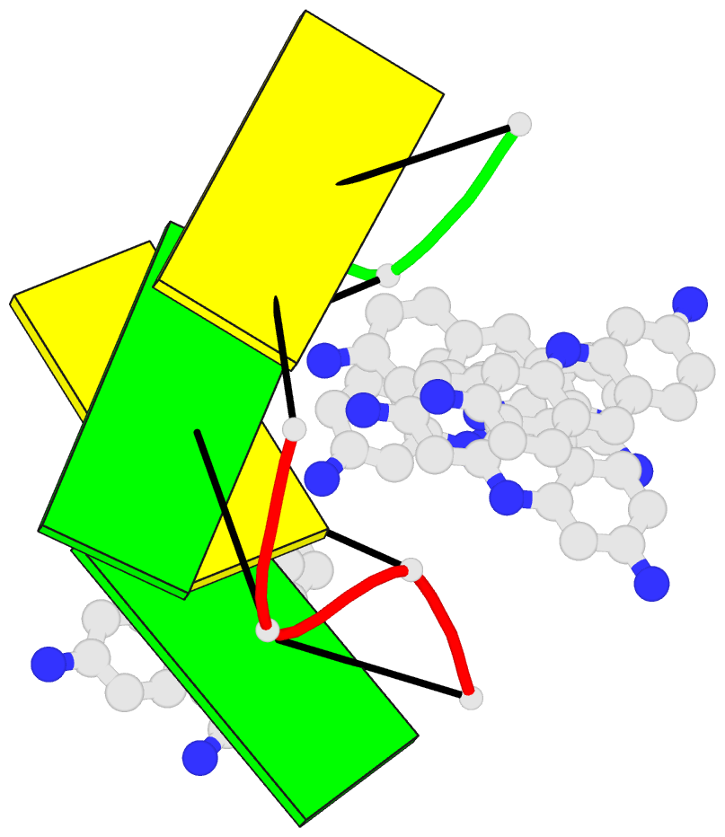
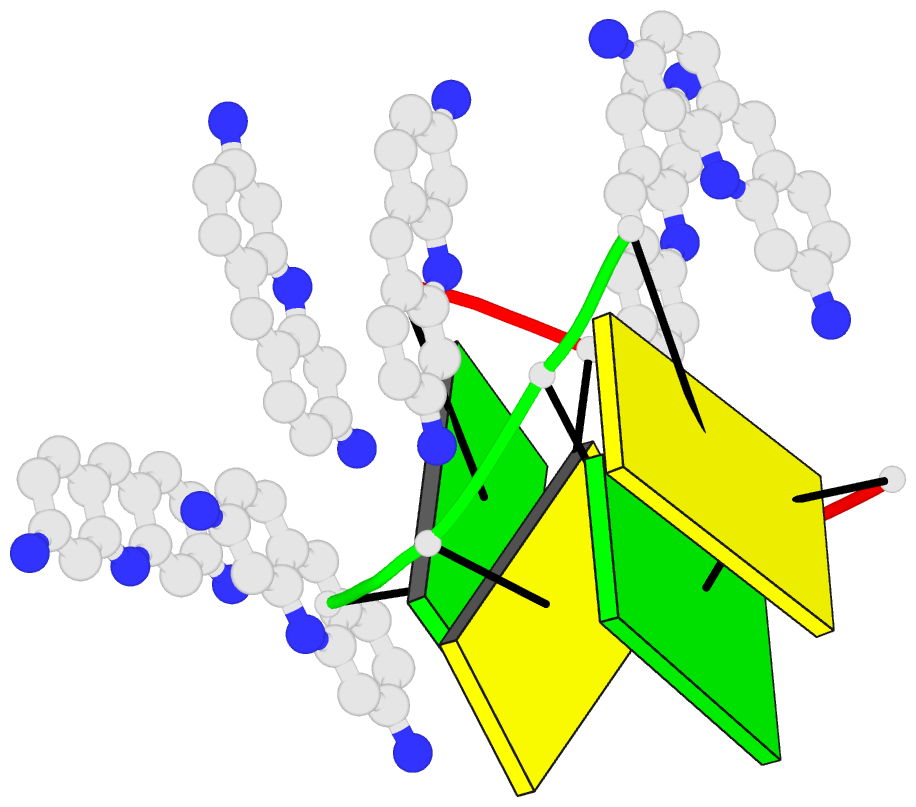
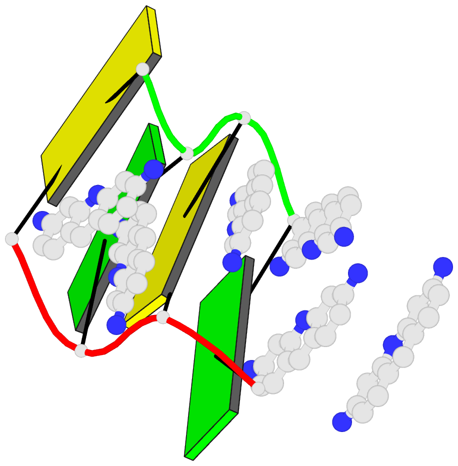
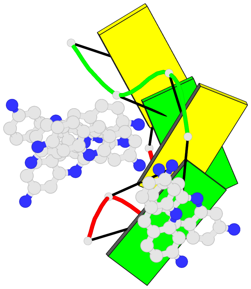
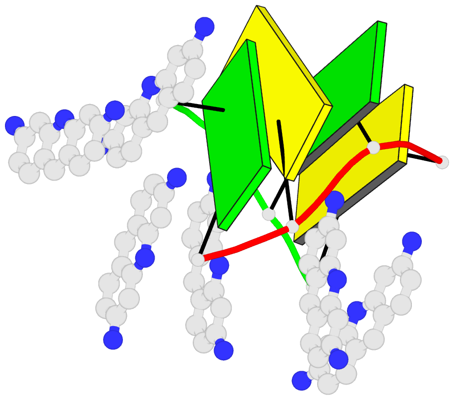
- Structure of 5'-d(*(bro)cp*gp*(bro)cp*g)-3' in complex with proflavine
- Reference
- Westhof E, Hosur MV, Sundaralingam M (1988): "Nonintercalative binding of proflavin to Z-DNA: structure of a complex between d(5BrC-G-5BrC-G) and proflavin." Biochemistry, 27, 5742-5747.
- Abstract
- The crystal structure of a disordered 1:1 complex between the tetradeoxyoligomer d(5BrC-G-5BrC-G) and proflavin has been determined and refined to an R factor of 26.9% for 474 reflections initially in space group P6(5) and to an R factor of 22.2% for 475 reflections in space group P2(1), both at 2-A resolution with Fobsd greater than or equal to 4.0. The unit cell constants are a = b = 17.9 A, c = 44.5 A, and gamma = 120 degrees. The final models are essentially the same in the two space groups with greater disorder in space group P6(5). In space group P2(1), the asymmetric unit is a tetranucleotide duplex, two sandwiched proflavin molecules, and four "outside-bound" proflavins. The tetranucleotide duplex is in the Z conformation and is located at the origin of the unit cell with a pair of proflavins sandwiched between the tetranucleotides. Thus, the tetranucleotides and proflavin dimers stack alternatively forming a quasi-continuous helix with the helix axis coincident with the c axis. The structure analysis revealed the presence of outside-bound proflavins as well. It is interesting that one type of outside-bound proflavins occupies a similar environment as the cobalt hexaammines in their complex with the decadeoxyoligomer d(CGTACGTACG) [Brennan, R. G., Westhof, E., & Sundaralingam, M. (1986) J. Biomol. Struct. Dyn. 3, 649]. Crystals of the latter are isomorphous to the present complex. The outside-bound proflavins penetrate the deep minor groove, thereby closing it off, and provide a visualization of a quasi-internal mode of binding of proflavin to a nucleic acid.