Summary information and primary citation
- PDB-id
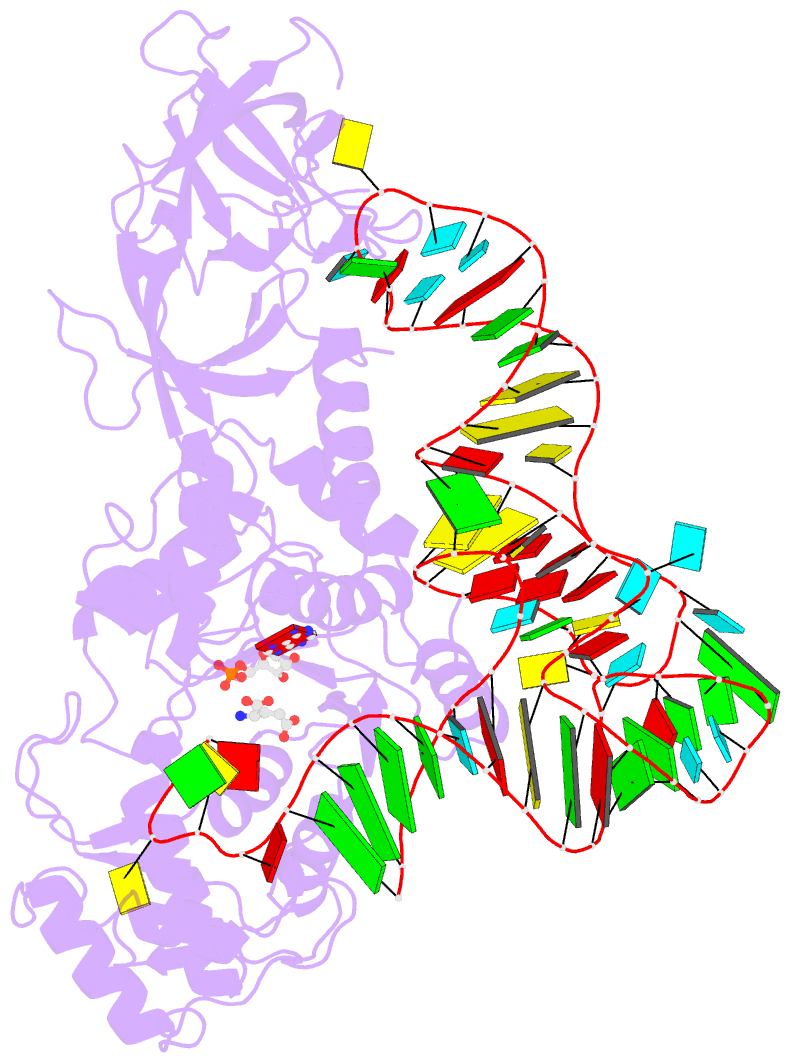
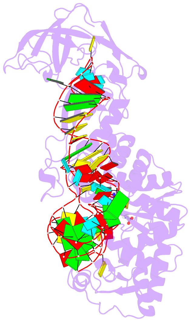
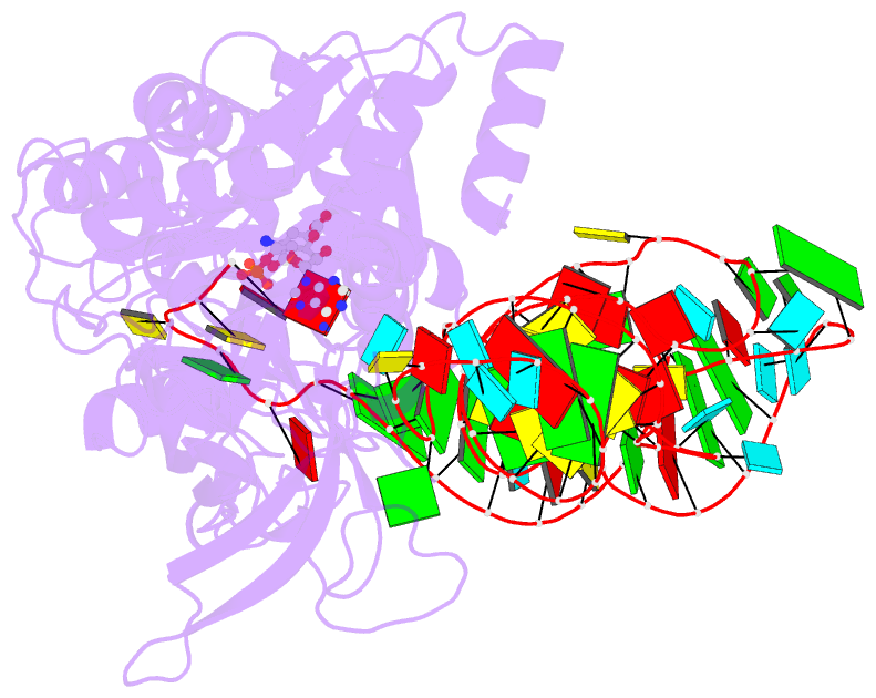
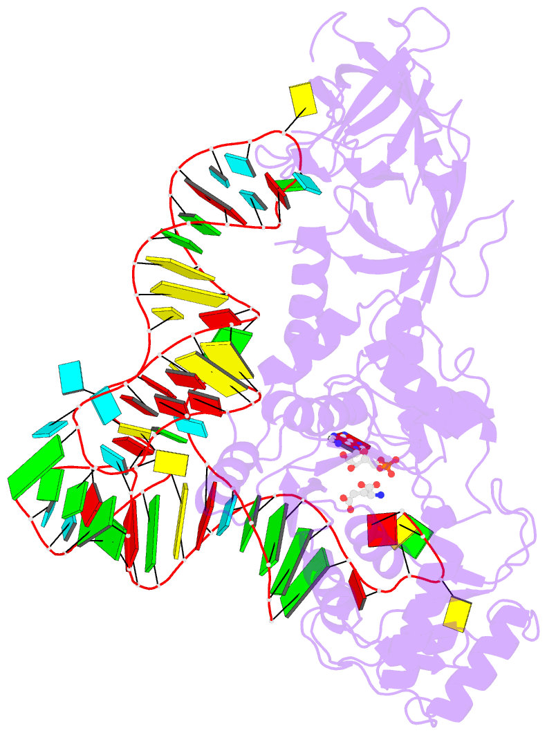
- 1o0c; DSSR-derived features in text and JSON formats
- Class
- ligase-RNA
- Method
- X-ray (2.7 Å)
- Summary
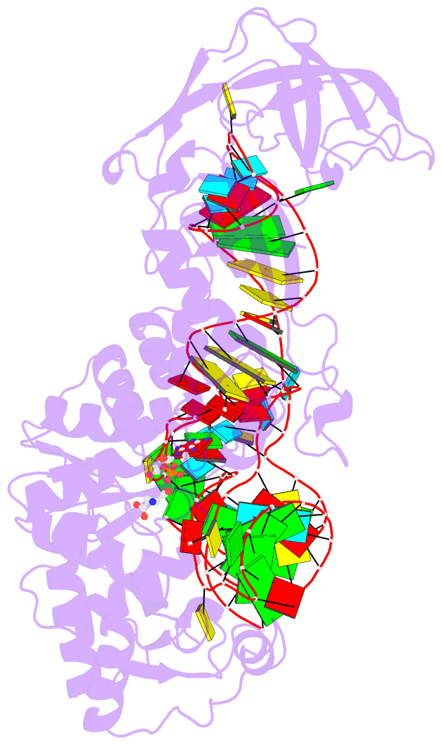
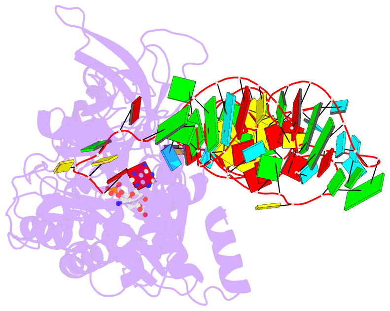
- Crystal structure of l-glutamate and ampcpp bound to glutamine aminoacyl trna synthetase
- Reference
- Bullock TL, Uter N, Nissan TA, Perona JJ (2003): "Amino Acid Discrimination by a class I aminoacyl-tRNA synthetase
specified by negative determinants." J.Mol.Biol., 328, 395-408. doi: 10.1016/S0022-2836(03)00305-X.
- Abstract
- The 2.5 A crystal structure of Escherichia coli glutaminyl-tRNA synthetase in a quaternary complex with tRNA(Gln), an ATP analog and glutamate reveals that the non-cognate amino acid adopts a distinct binding mode within the active site cleft. In contrast to the binding of cognate glutamine, one oxygen of the charged glutamate carboxylate group makes a direct ion-pair interaction with the strictly conserved Arg30 residue located in the first half of the dinucleotide fold domain. The nucleophilic alpha-carboxylate moiety of glutamate is mispositioned with respect to both the ATP alpha-phosphate and terminal tRNA ribose groups, suggesting that a component of amino acid discrimination resides at the catalytic step of the reaction. Further, the other side-chain carboxylate oxygen of glutamate is found in a position identical to that previously proposed to be occupied by the NH(2) group of the cognate glutamine substrate. At this position, the glutamate oxygen accepts hydrogen bonds from the hydroxyl moiety of Tyr211 and a water molecule. These findings demonstrate that amino acid specificity by GlnRS cannot arise from hydrogen bonds donated by the cognate glutamine amide to these same moieties, as previously suggested. Instead, Arg30 functions as a negative determinant to drive binding of non-cognate glutamate into a non-productive orientation. The poorly differentiated cognate amino acid-binding site in GlnRS may be a consequence of the late emergence of this enzyme from the eukaryotic lineage of glutamyl-tRNA synthetases.