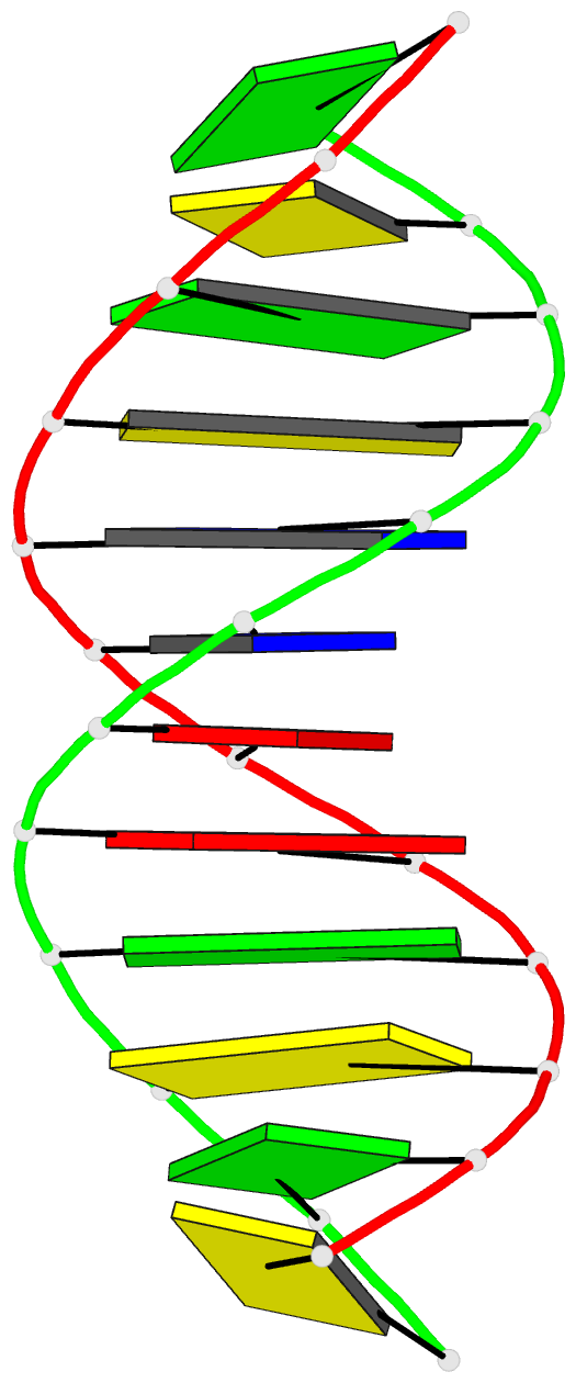
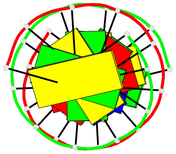
Summary information and primary citation
- PDB-id
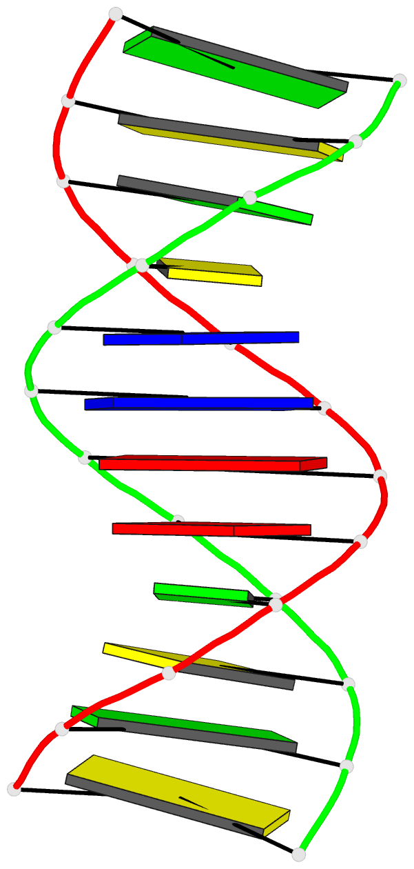
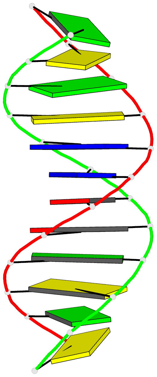
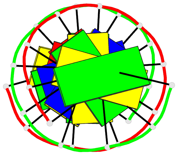
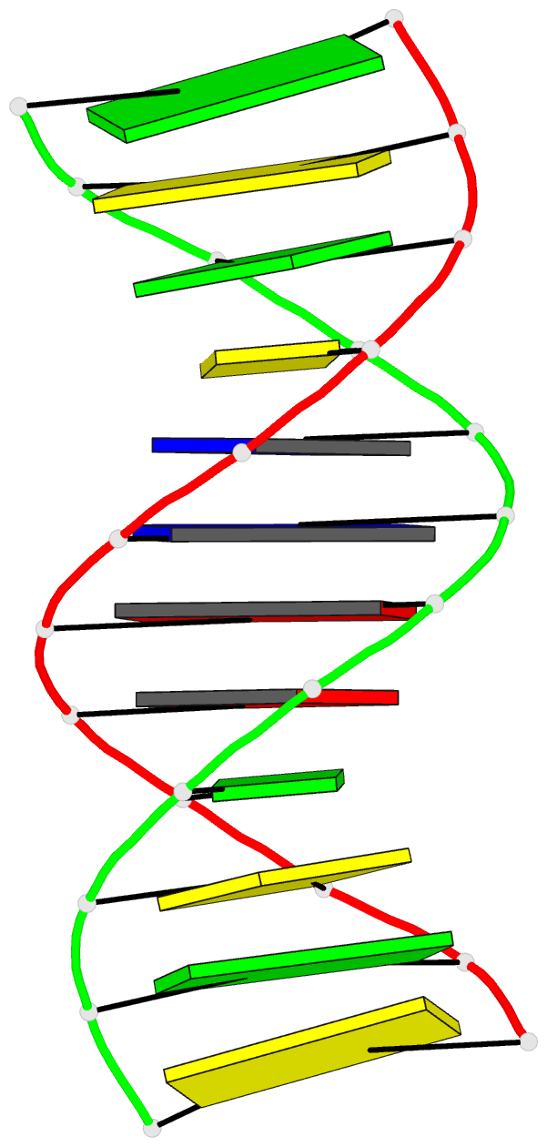
- 1naj; DSSR-derived features in text and JSON formats
- Class
- DNA
- Method
- NMR
- Summary
- High resolution NMR structure of DNA dodecamer determined in aqueous dilute liquid crystalline phase
- Reference
- Wu Z, Delaglio F, Tjandra N, Zhurkin VB, Bax A (2003): "Overall structure and sugar dynamics of a DNA dodecamer from homo- and heteronuclear dipolar couplings and (31)P chemical shift anisotropy." J.Biomol.Nmr, 26, 297-315. doi: 10.1023/A:1024047103398.
- Abstract
- The solution structure of d(CGCGAATTCGCG)(2) has been determined on the basis of an exceptionally large set of residual dipolar couplings. In addition to the heteronuclear (13)C-(1)H and (15)N-(1)H and qualitative homonuclear (1)H-(1)H dipolar couplings, previously measured in bicelle medium, more than 300 quantitative (1)H-(1)H and 22 (31)P-(1)H dipolar restraints were obtained in liquid crystalline Pf1 medium, and 22 (31)P chemical shift anisotropy restraints. High quality DNA structures can be obtained solely on the basis of these new restraints, and these structures are in close agreement with those calculated previously on the basis of (13)C-(1)H and (15)N-(1)H dipolar couplings. In the newly calculated structures, (31)P-(1)H dipolar and (3)JsubH3(')Psub couplings and (31)P CSA data restrain the phosphodiester backbone torsion angles. The final structure represents a quite regular B-form helix with a modest bending of approximately 10 degrees, which is essentially independent of whether or not electrostatic terms are used in the calculation. Combined, the number of homo- and heteronuclear dipolar couplings significantly exceeds the number of degrees of freedom in the system. Results indicate that the dipolar coupling data cannot be fit by a single structure, but are compatible with the presence of rapid equilibria between C2(')-endo and C3(')-endo deoxyribose puckers (sugar switching). The C2(')-H2(')/H2(") dipolar couplings in B-form DNA are particularly sensitive to sugar pucker and yield the largest discrepancies when fit to a single structure. To resolve these discrepancies, we suggest a simplified dipolar coupling analysis that yields N/S equilibria for the ribose sugar puckers, which are in good agreement with previous analyses of NMR J(HH) couplings, with a population of the minor C3(')-endo form higher for pyrimidines than for purines.