Summary information and primary citation
- PDB-id
- 1kpy; DSSR-derived features in text and JSON formats
- Class
- RNA
- Method
- NMR
- Summary
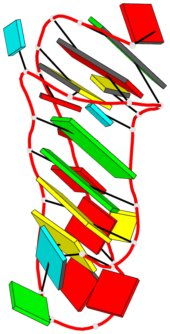
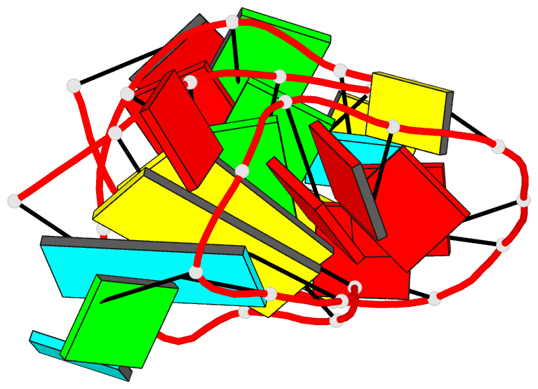
- Pemv-1 p1-p2 frameshifting pseudoknot, 15 lowest energy structures
- Reference
- Nixon PL, Rangan A, Kim Y-G, Rich A, Hoffman DW, Hennig M, Giedroc DP (2002): "Solution structure of a luteoviral P1-P2 frameshifting mRNA pseudoknot." J.Mol.Biol., 322, 621-633. doi: 10.1016/S0022-2836(02)00779-9.
- Abstract
- A hairpin-type messenger RNA pseudoknot from pea enation mosaic virus RNA1 (PEMV-1) regulates the efficiency of programmed -1 ribosomal frameshifting. The solution structure and 15N relaxation rates reveal that the PEMV-1 pseudoknot is a compact-folded structure composed almost entirely of RNA triple helix. A three nucleotide reverse turn in loop 1 positions a protonated cytidine, C(10), in the correct orientation to form an A((n-1)).C(+).G-C(n) major groove base quadruple, like that found in the beet western yellows virus pseudoknot and the hepatitis delta virus ribozyme, despite distinct structural contexts. A novel loop 2-loop 1 A.U Hoogsteen base-pair stacks on the C(10)(+).G(28) base-pair of the A(12).C(10)(+).G(28)-C(13) quadruple and forms a wedge between the pseudoknot stems stabilizing a bent and over-rotated global conformation. Substitution of key nucleotides that stabilize the unique conformation of the PEMV-1 pseudoknot greatly reduces ribosomal frameshifting efficacy.