Summary information and primary citation
- PDB-id
- 1k1h; DSSR-derived features in text and JSON formats
- Class
- DNA
- Method
- NMR
- Summary
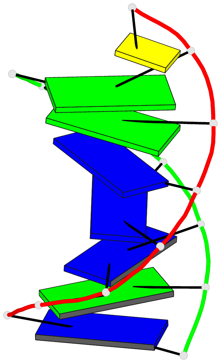
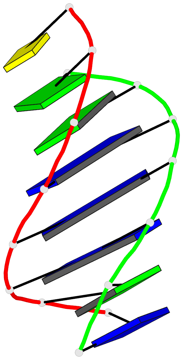
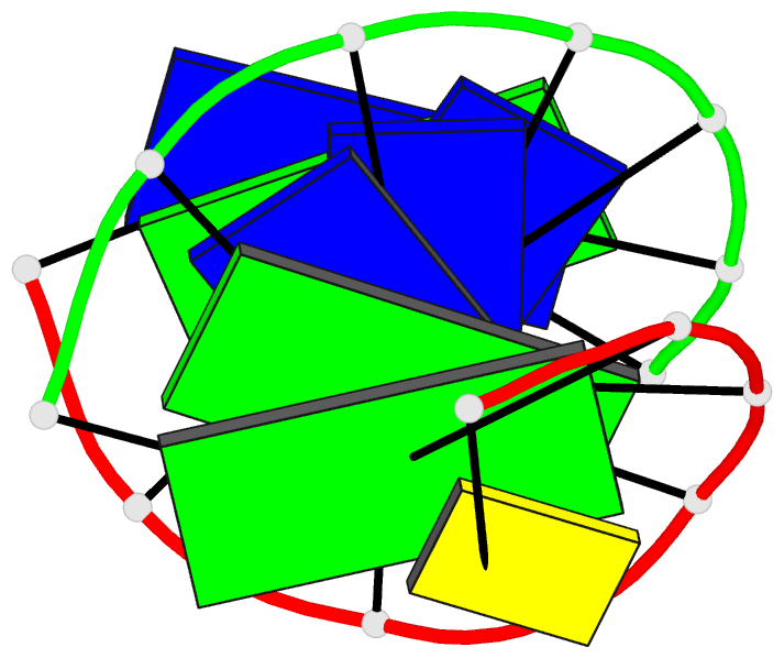
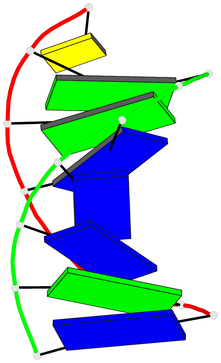
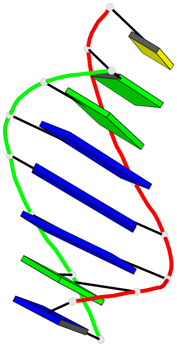
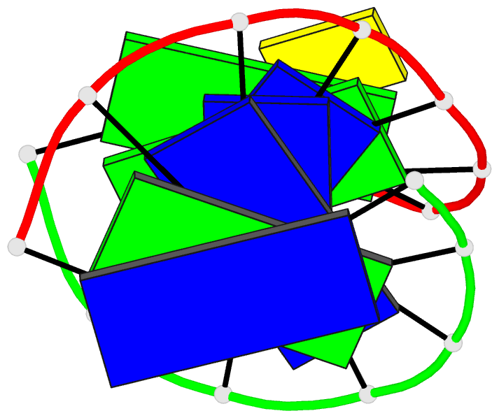
- Heteroduplex of chirally pure methylphosphonate-DNA duplex
- Reference
- Thiviyanathan V, Vyazovkina KV, Gozansky EK, Bichenchova E, Abramova TV, Luxon BA, Lebedev AV, Gorenstein DG (2002): "Structure of hybrid backbone methylphosphonate DNA heteroduplexes: effect of R and S stereochemistry." Biochemistry, 41, 827-838. doi: 10.1021/bi011551k.
- Abstract
- Methyl phosphonate oligonucleotides have been used as antisense and antigene agents. Substitution of a methyl group for oxygen in the phosphate ester backbone introduces a new chiral center. Significant differences in physical properties and hybridization abilities are observed between the R(p) and S(p) diastereomers. Chirally pure methylphosphonate deoxyribooligonucleotides were synthesized, and the solution structures of duplexes formed between a single strand heptanucleotide methylphosphonate, d(Cp(Me)Cp(Me)Ap(Me)Ap(Me)Ap(Me)Cp(Me)A), hybridized to a complementary octanucleotide, d(TpGpTpTpTpGpGpC), were studied by NMR spectroscopy. Stereochemistry at the methylphosphonate center for the heptanucleotide was either RpRpRpRpRpRp (R(p) stereoisomer) or RpRpRpSpRpRp (S(p) stereoisomer, although only one of the six methylphosphonate centers has the S(p) stereochemistry). The results show that the methylphosphonate strands in the heteroduplexes exhibit increased dynamics relative to the DNA strand. Substitution of one chiral center from R(p) to S(p) has a profound effect on the hybridization ability of the methylphosphonate strand. Sugars in the phosphodiester strand exhibit C(2)(') endo sugar puckering while the sugars in the methyl phosphonate strand exhibit an intermediate C(4)(') endo puckering. Bases are well stacked on each other throughout the duplex. The hybridization of the methylphosphonate strand does not perturb the structure of the complementary DNA strand in the hetero duplexes. The sugar residue 5' to the S(p) chiral center shows A-form sugar puckering, with a C(3)(')-endo conformation. Minor groove width in the R(p) stereoisomer is considerably wider, particularly at the R(p) vs S(p) site and is attributed to either steric interactions across the minor groove or poorer metal ion coordination within the minor groove.