Summary information and primary citation
- PDB-id
- 1i6u; DSSR-derived features in text and JSON formats
- Class
- ribosome
- Method
- X-ray (2.6 Å)
- Summary
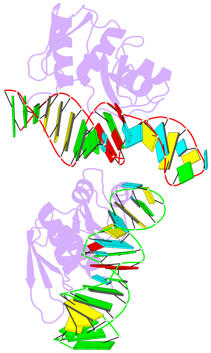
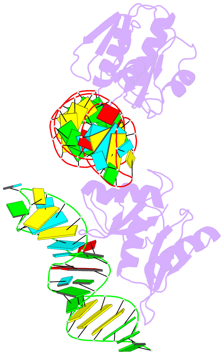
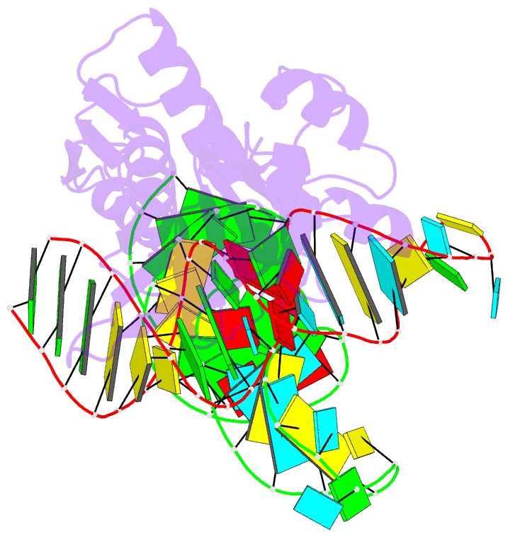
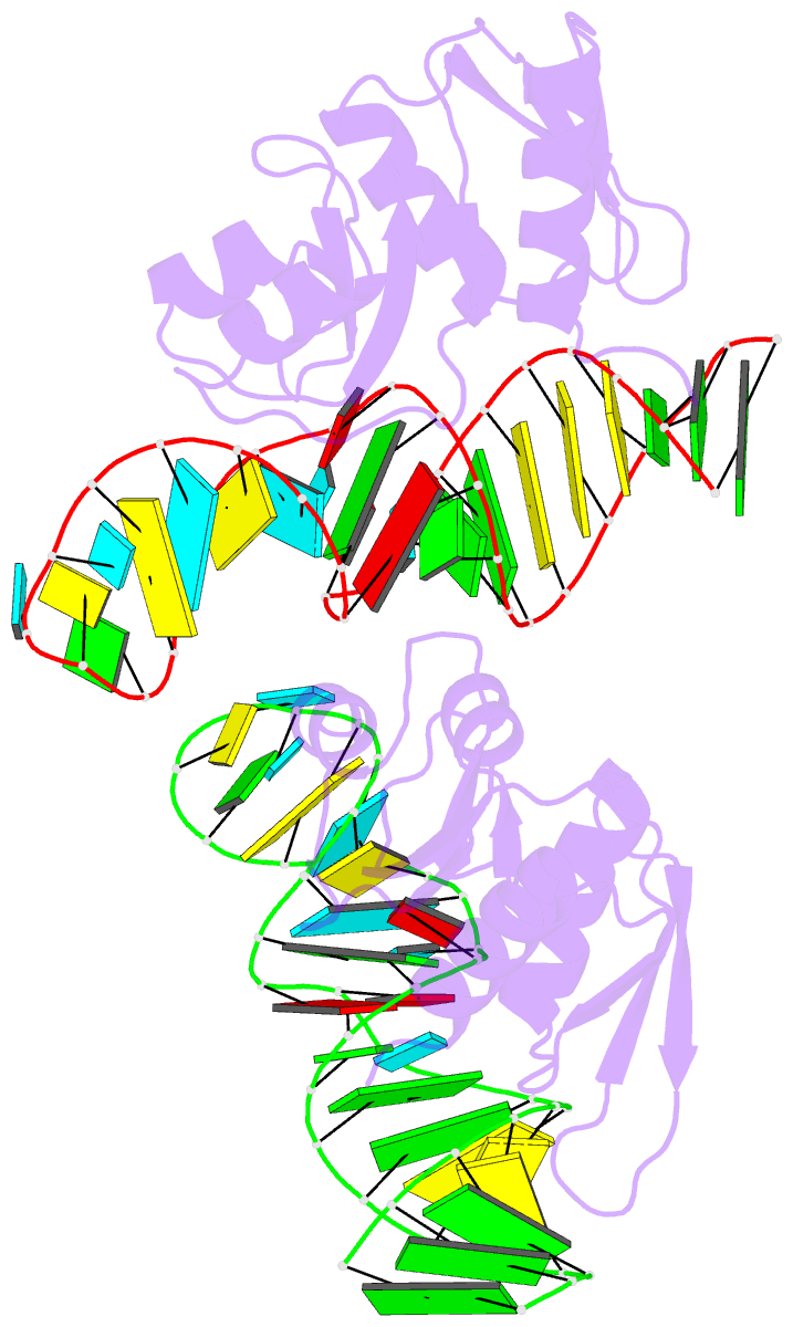
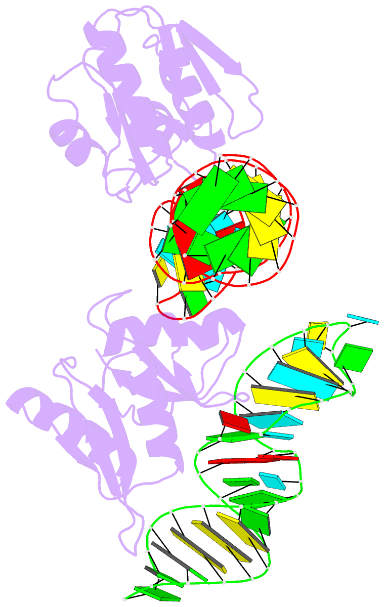
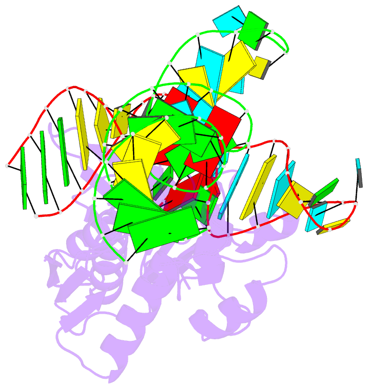
- RNA-protein interactions: the crystal structure of ribosomal protein s8-rrna complex from methanococcus jannaschii
- Reference
- Tishchenko S, Nikulin A, Fomenkova N, Nevskaya N, Nikonov O, Dumas P, Moine H, Ehresmann B, Ehresmann C, Piendl W, Lamzin V, Garber M, Nikonov S (2001): "Detailed analysis of RNA-protein interactions within the ribosomal protein S8-rRNA complex from the archaeon Methanococcus jannaschii." J.Mol.Biol., 311, 311-324. doi: 10.1006/jmbi.2001.4877.
- Abstract
- The crystal structure of ribosomal protein S8 bound to its target 16 S rRNA from a hyperthermophilic archaeon Methanococcus jannaschii has been determined at 2.6 A resolution. The protein interacts with the minor groove of helix H21 at two sites located one helical turn apart, with S8 forming a bridge over the RNA major groove. The specificity of binding is essentially provided by the C-terminal domain of S8 and the highly conserved nucleotide core, characterized by two dinucleotide platforms, facing each other. The first platform (A595-A596), which is the less phylogenetically and structurally constrained, does not directly contact the protein but has an important shaping role in inducing cross-strand stacking interactions. The second platform (U641-A642) is specifically recognized by the protein. The universally conserved A642 plays a pivotal role by ensuring the cohesion of the complex organization of the core through an array of hydrogen bonds, including the G597-C643-U641 base triple. In addition, A642 provides the unique base-specific interaction with the conserved Ser105, while the Thr106 - Thr107 peptide link is stacked on its purine ring. Noteworthy, the specific recognition of this tripeptide (Thr-Ser-Thr/Ser) is parallel to the recognition of an RNA tetraloop by a dinucleotide platform in the P4-P6 ribozyme domain of group I intron. This suggests a general dual role of dinucleotide platforms in recognition of RNA or peptide motifs. One prominent feature is that conserved side-chain amino acids, as well as conserved bases, are essentially involved in maintaining tertiary folds. The specificity of binding is mainly driven by shape complementarity, which is increased by the hydrophobic part of side-chains. The remarkable similarity of this complex with its homologue in the T. thermophilus 30 S subunit indicates a conserved interaction mode between Archaea and Bacteria.