Summary information and primary citation
- PDB-id
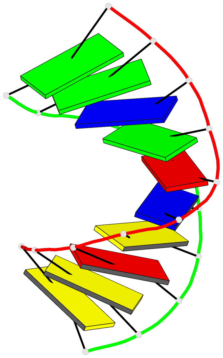
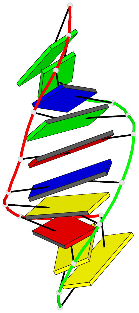
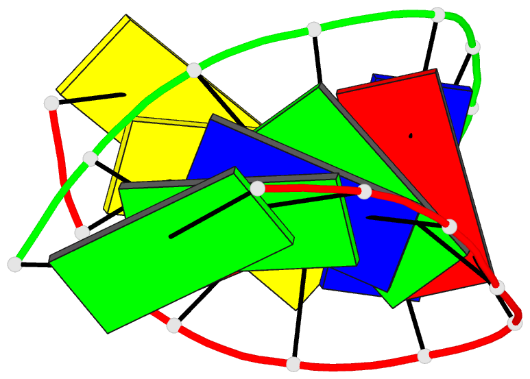
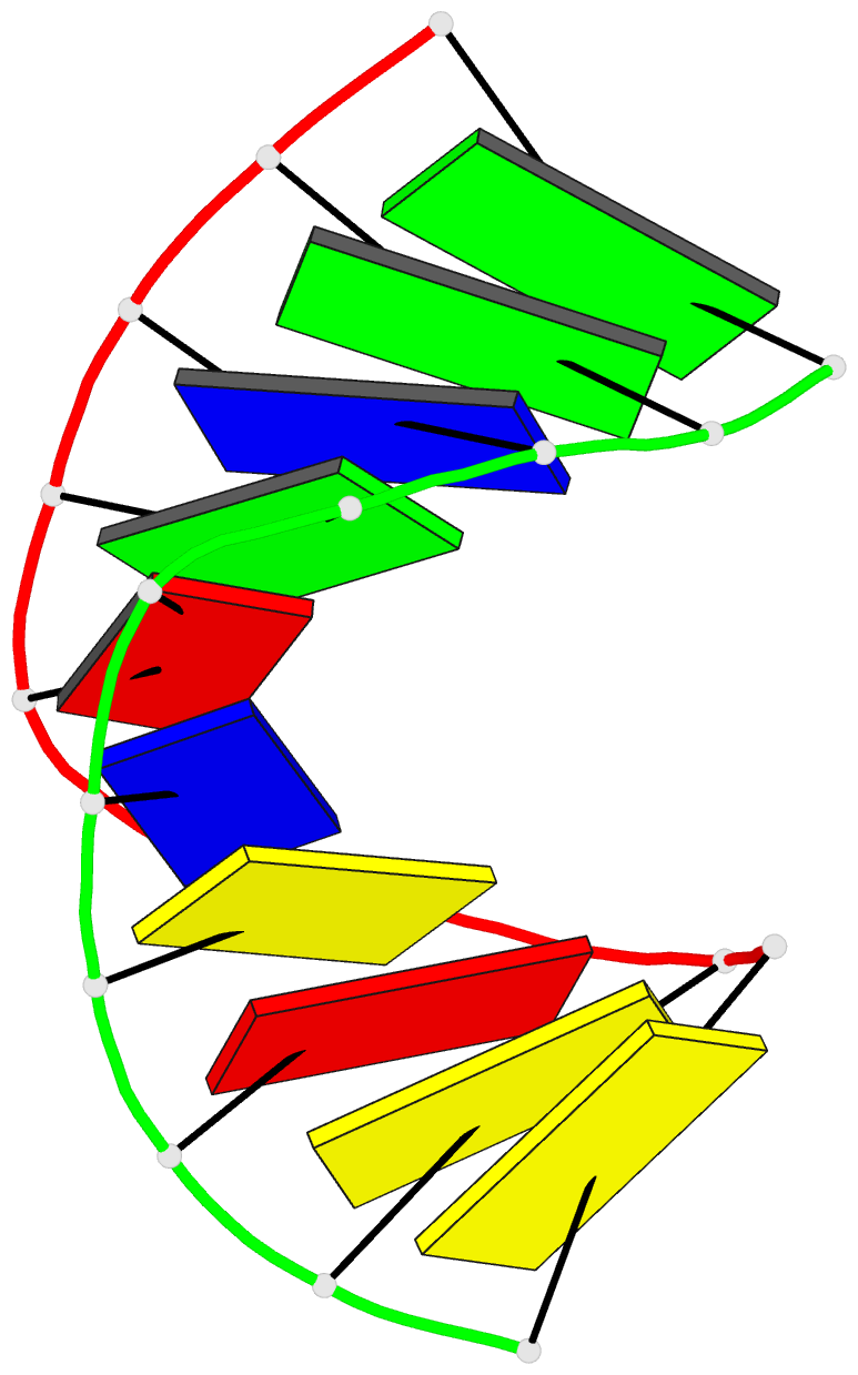
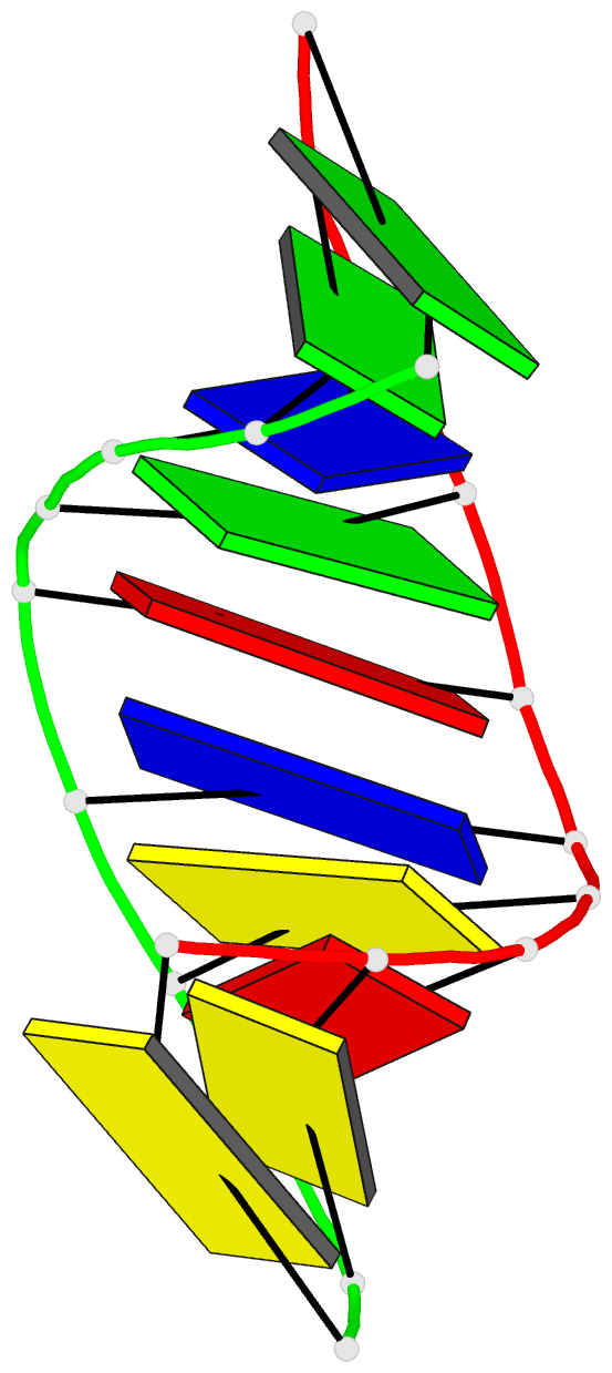
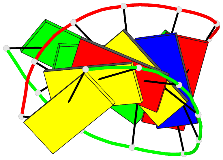
- 1evp; DSSR-derived features in text and JSON formats
- Class
- DNA-RNA hybrid
- Method
- X-ray (1.8 Å)
- Summary
- Crystal structure of the chimerical decamer d(ccactagtg)r(g)
- Reference
- Wahl MC, Sundaralingam M (2000): "B-form to A-form conversion by a 3'-terminal ribose: crystal structure of the chimera d(CCACTAGTG)r(G)." Nucleic Acids Res., 28, 4356-4363. doi: 10.1093/nar/28.21.4356.
- Abstract
- The crystal structure of the chimerical decamer d(CCACTAGTG)r(G), bearing a 3'-terminal ribo-guanidine, has been solved and refined at 1.8 A resolution (R-factor 16.6%; free R-factor 22.8%). The decamer crystallizes in the orthorhombic space group P2(1)2(1)2(1) with unit cell constants a = 23.90 A, b = 45.76 A and c = 49.27 A. The structure was solved by molecular replacement using the coordinates of the isomorphous chimera r(GCG)d(TATACGC). The final model contains one duplex and 77 water molecules per asymmetric unit. Surprisingly, all residues adopt a conformation typical for A-form nucleic acids (C3'-endo type sugar pucker) although the all-DNA analog, d(CCACTAGTGG), has been crystallized in the B-form. Comparing circular dichroism spectra of the chimera and the corresponding all-DNA sequence reveals a similar trend of the former molecule to adopt an A-like conformation in solution. The results suggest that the preference of ribonucleotides for the A-form is communicated into the 5'-direction of an oligonucleotide strand, although direct interactions of the 2'-hydroxyl group can only be discerned with nucleotides in the 3'-direction of a C3'-endo puckered ribose. These observations imply that forces like water-mediated contacts, the concerted motions of backbone torsion angles, and stacking preferences, are responsible for such long-range influences. This bi-directional structural communication originating from a ribonucleotide can be expected to contribute to the stability of the A-form within all-RNA duplexes.