Summary information and primary citation
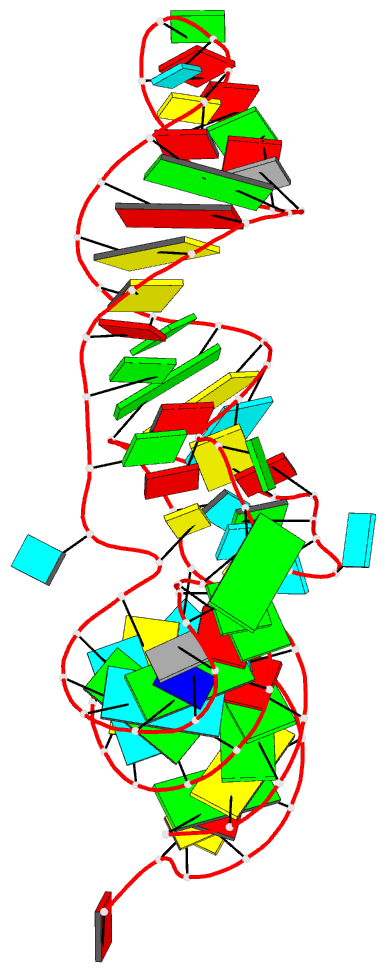
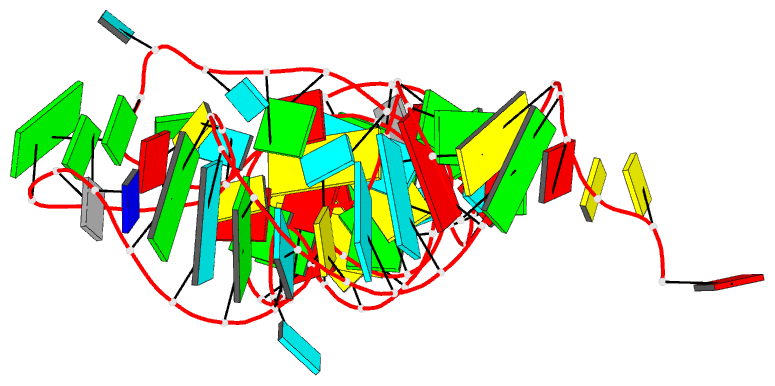
- PDB-id
-
1ehz;
SNAP-derived features in text and
JSON formats
- Class
- RNA
- Method
- X-ray (1.93 Å)
- Summary
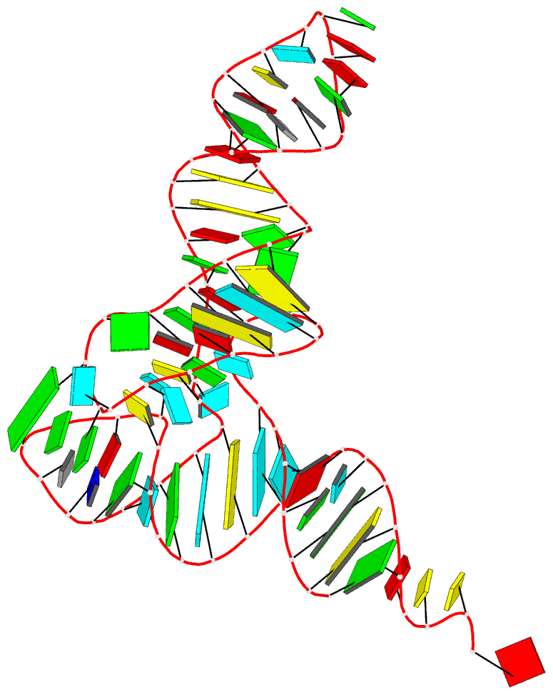
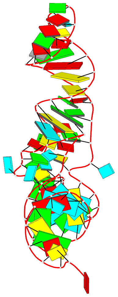
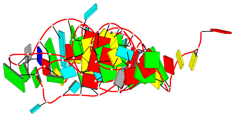
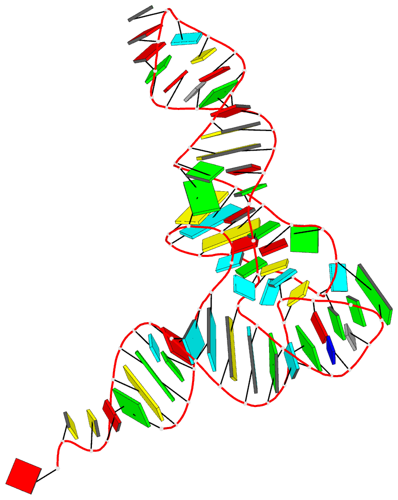
- The crystal structure of yeast phenylalanine trna at
1.93 Å resolution
- Reference
-
Shi H, Moore PB (2000): "The
crystal structure of yeast phenylalanine tRNA at 1.93 A
resolution: a classic structure revisited."
RNA, 6, 1091-1105. doi:
10.1017/S1355838200000364.
- Abstract
- The crystal structure of the monoclinic form of yeast
phenylalanine tRNA has been redetermined at a resolution of
1.93 A. The structure of yeast tRNAphe described here is
more accurate than its predecessors not only because it
incorporates higher resolution data, but also because it
has been refined using techniques that had not been
developed when its predecessors were determined more than
20 years ago. The 1.93 A resolution version of this
structure differs interestingly from its predecessors in
its details. In loop regions particularly, the backbone
torsion angles in the new structure are not the same as
those reported earlier. Several new divalent cation binding
sites have been identified, and the water structure that
has emerged is also different.