Summary information and primary citation
- PDB-id
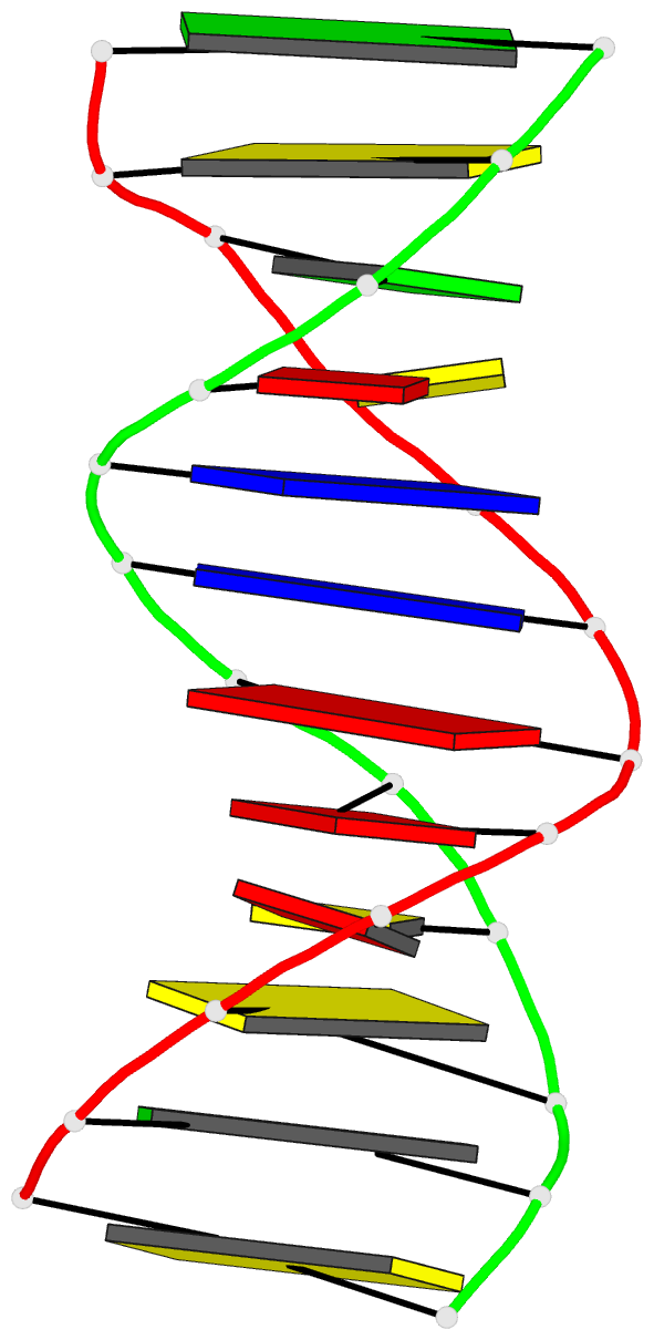
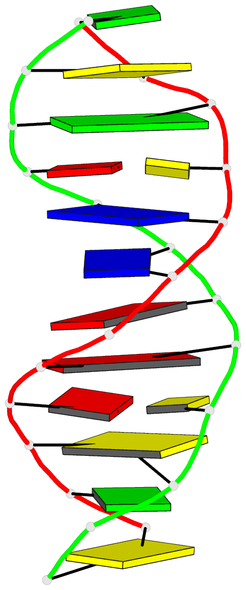
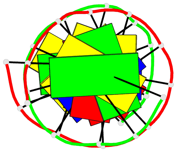
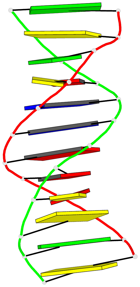
- 1d99; DSSR-derived features in text and JSON formats
- Class
- DNA
- Method
- X-ray (2.5 Å)
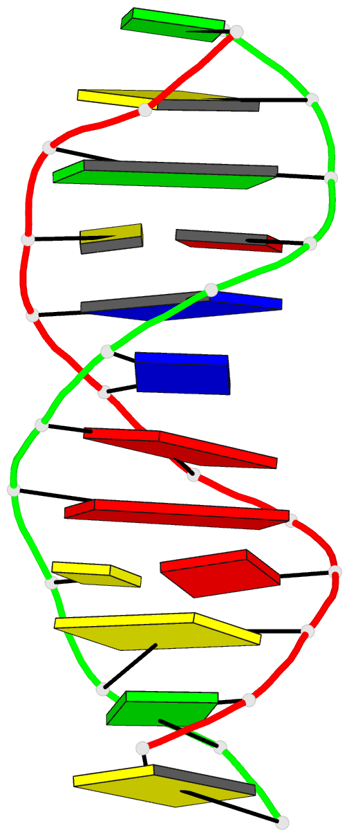
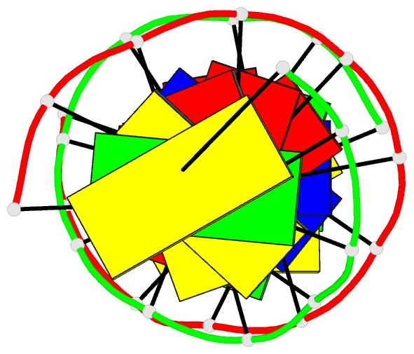
- Summary
- Structural features and hydration of a dodecamer duplex containing two c.a mispairs
- Reference
- Hunter WN, Brown T, Kennard O (1987): "Structural features and hydration of a dodecamer duplex containing two C.A mispairs." Nucleic Acids Res., 15, 6589-6605. doi: 10.1093/nar/15.16.6589.
- Abstract
- X-ray diffraction techniques have been used to characterise the crystal and molecular structure of the deoxyoligomer d(C-G-C-A-A-A-T-T-C-G-C-G) at 2.5A resolution. The final R factor is 0.19 with the location of 78 solvent molecules. The oligomer crystallises in a B-DNA type conformation with two strands coiled about each other to produce a duplex. This double helix consists of four A.T and six G.C Watson-Crick base pairs and two C.A mispairs. The mismatched base pairs adopt a "wobble" type structure with the cytosine displaced laterally into the major groove, the adenine into the minor groove. We have proposed that the two close contacts observed in the C.A pairing represent two hydrogen bonds one of which results from protonation of adenine. The mispairs are accommodated in the double helix with small adjustments in the conformation of the sugar-phosphate backbone. Details of the backbone conformation, base stacking interactions, thermal parameters and the hydration are now presented and compared with those of the native oligomer d(C-G-C-G-A-A-T-T-C-G-C-G) and with variations of this sequence containing G.T and G.A mispairs.