Summary information and primary citation
- PDB-id
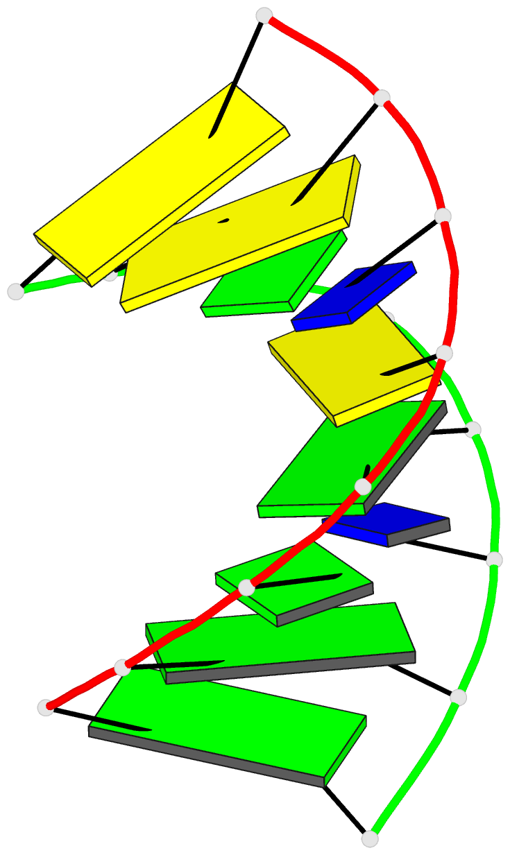
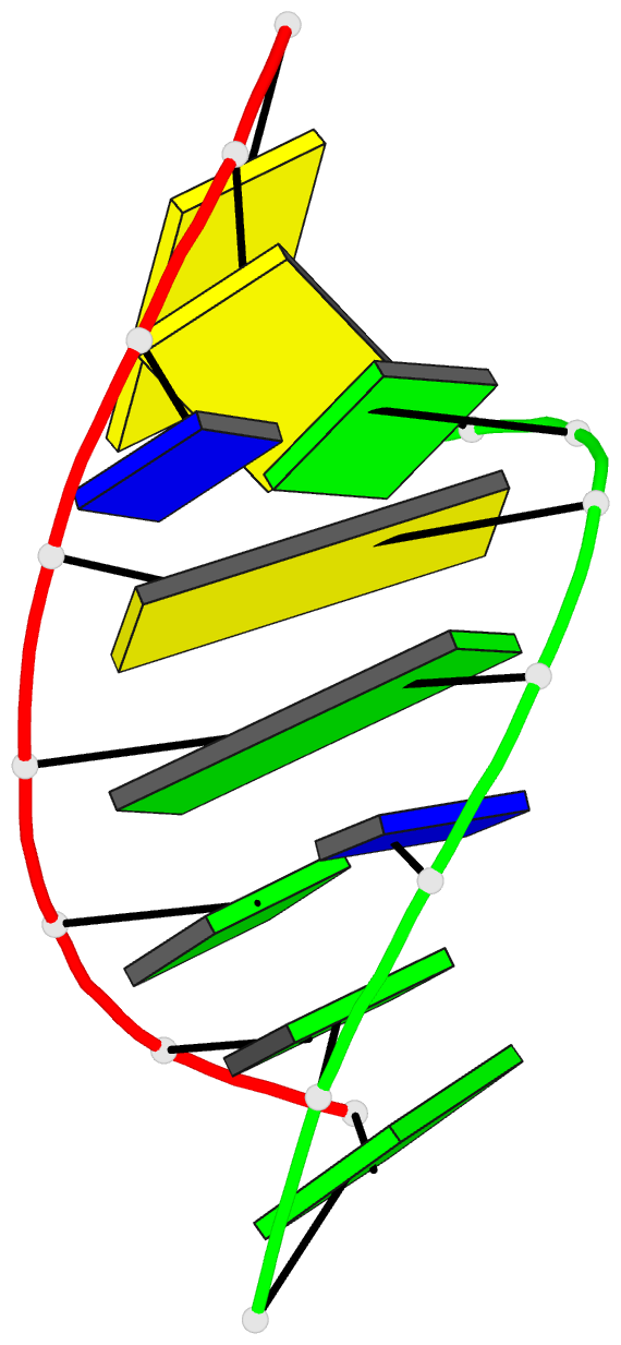
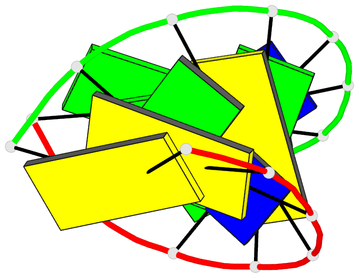
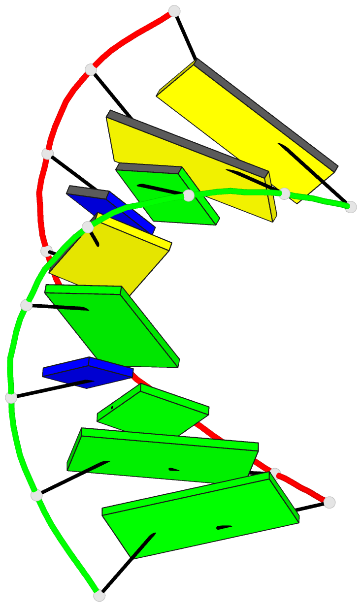
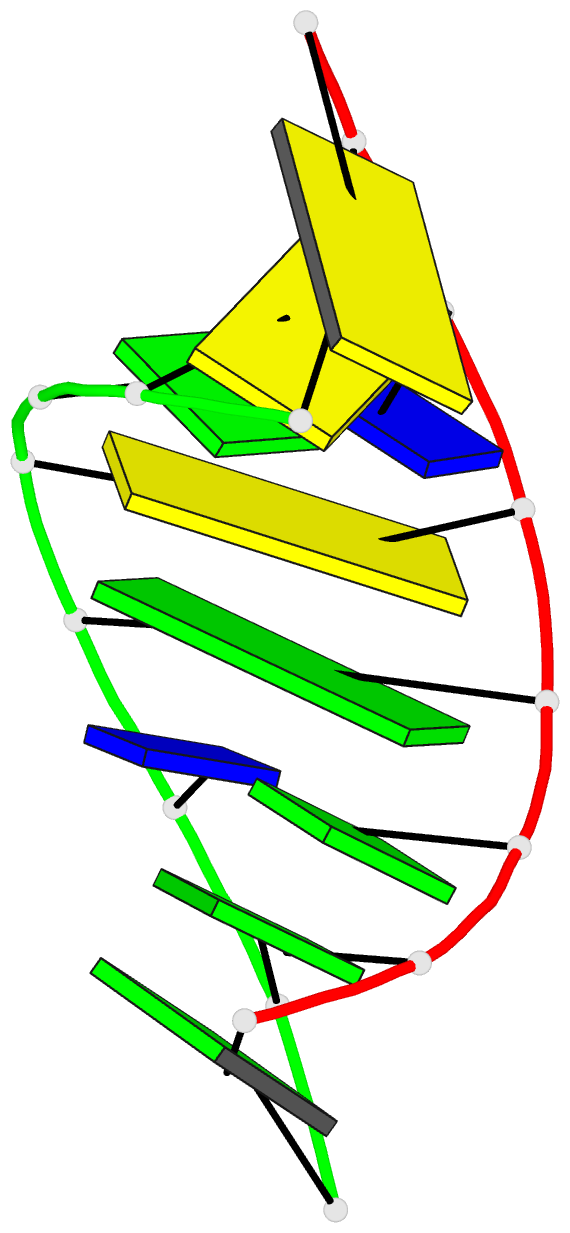
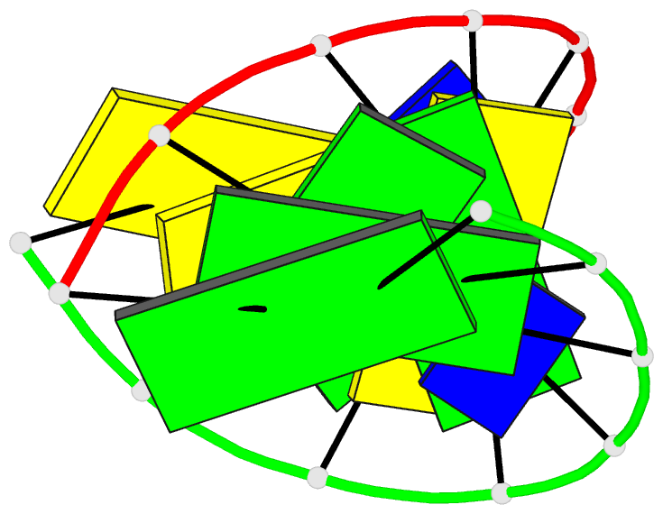
- 1d90; DSSR-derived features in text and JSON formats
- Class
- DNA
- Method
- X-ray (1.7 Å)
- Summary
- Refined crystal structure of an octanucleotide duplex with i.t mismatched base pairs
- Reference
- Cruse WB, Aymani J, Kennard O, Brown T, Jack AG, Leonard GA (1989): "Refined crystal structure of an octanucleotide duplex with I.T. mismatched base pairs." Nucleic Acids Res., 17, 55-72. doi: 10.1093/nar/17.1.55.
- Abstract
- The structure of the synthetic deoxyoctamer d(GGIGCTCC) has been determined by single crystal X-ray diffraction techniques to a resolution of 1.7A. The sequence crystallises in space group P6(1), with unit cell dimensions a = b = 45.07, c = 45.49A. The refinement converged with a crystallographic residual R = 0.14 and the location of 81 solvent molecules. The octamer forms an A-DNA duplex with 6 Watson-Crick (G.C) base pairs and 2 inosine-thymine (I.T) pairs. Refinement of the structure shows it to be essentially isomorphous with that reported for d(GGGGCTCC) with the mispairs adopting a "wobble" conformation. Conformational parameters and base stacking interactions are compared to those for the native duplex d(GGGGCCCC) and other similar sequences. A rationale for the apparent increased crystal packing efficiency and lattice stability of the I.T octamer is given.