Summary information and primary citation
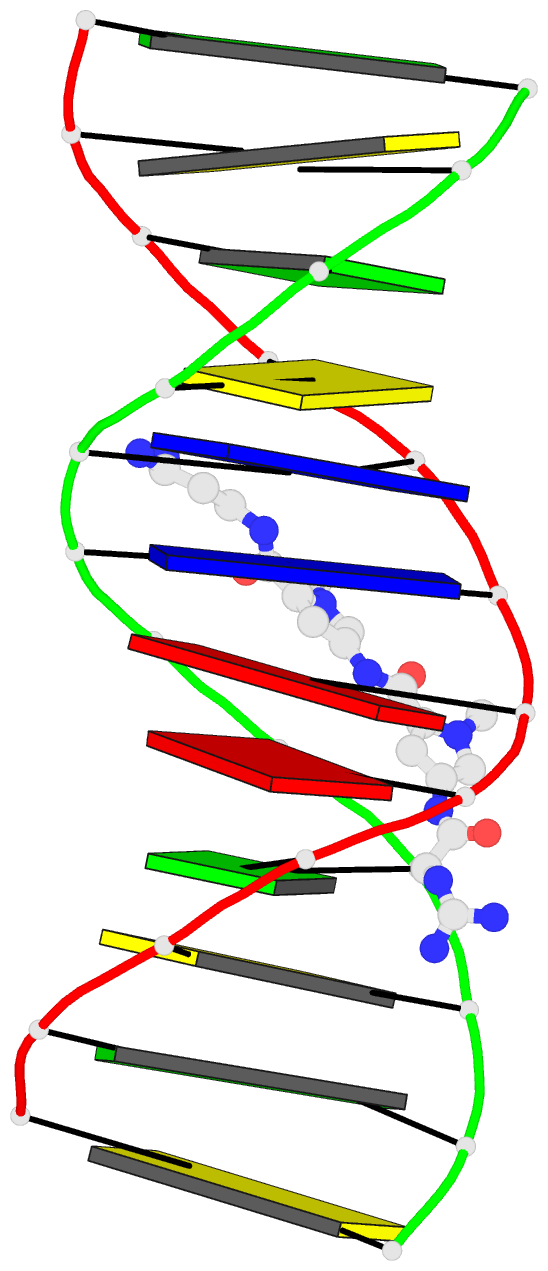
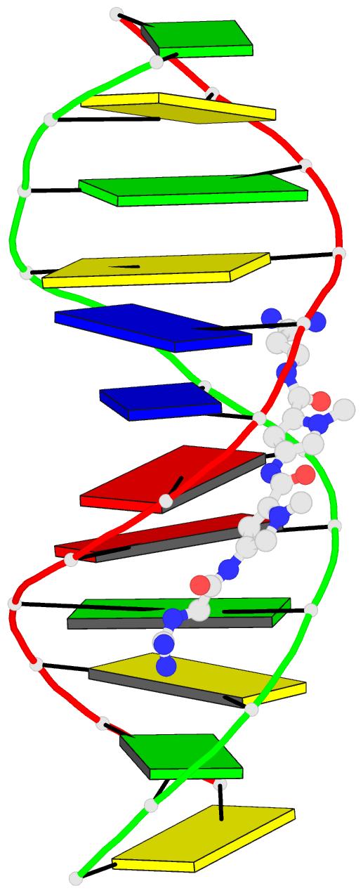
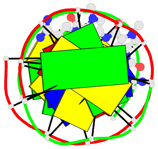
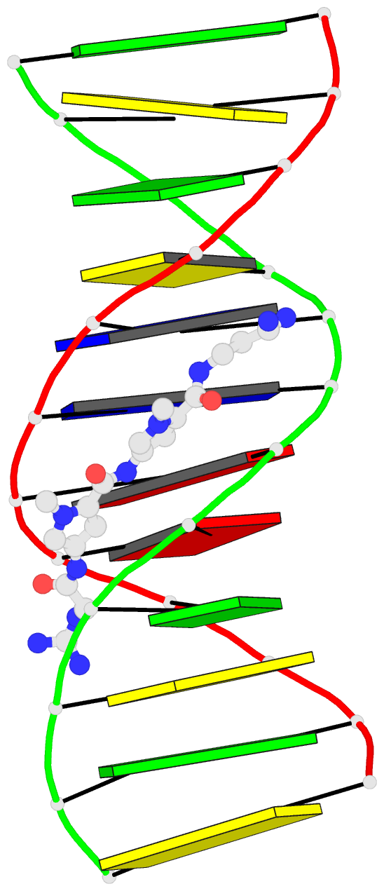
- PDB-id
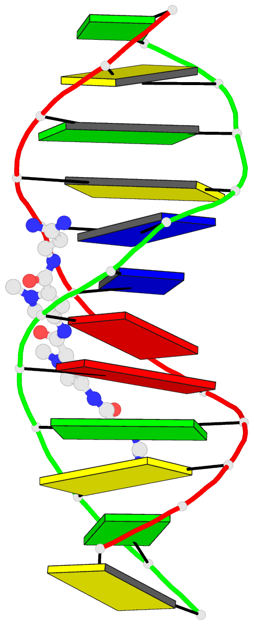
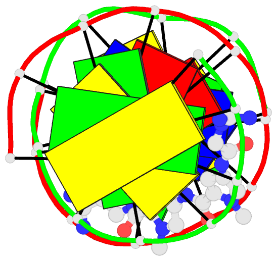
- 1d85; DSSR-derived features in text and JSON formats
- Class
- DNA
- Method
- X-ray (2.5 Å)
- Summary
- Structural consequences of a carcinogenic alkylation lesion on DNA: effect of o6-ethyl-guanine on the molecular structure of d(cgc[e6g]aattcgcg)-netropsin complex
- Reference
- Sriram M, van der Marel GA, Roelen HL, van Boom JH, Wang AH (1992): "Structural consequences of a carcinogenic alkylation lesion on DNA: effect of O6-ethylguanine on the molecular structure of the d(CGC[e6G]AATTCGCG)-netropsin complex." Biochemistry, 31, 11823-11834. doi: 10.1021/bi00162a022.
- Abstract
- Exposure of cells to alkylating agents produces DNA lesions, most of which are repaired. However some alkyl lesions persist and play a role in inducing point mutations and the subsequent carcinogenic conversion. O6-Ethylguanine (e6G) is a relatively persistent alkylation lesion caused by the exposure of DNA to N-ethyl-N-nitrosourea. We study the consequence of the e6G incorporation in DNA by X-ray crystallography. We have obtained crystals of the modified DNA dodecamer d(CGC[e6G]AATTCGCG) and the unmodified d(CGCGAATTCGCG), complexed to the minor groove binding drug netropsin. The space group of both crystals is P2(1)2(1)2(1), isomorphous to other related dodecamer DNA crystals. The structures have been solved by the molecular replacement method and refined by the constrained least-squares procedure to R-factors of approximately 16% at resolution of approximately 2.5 A. The two independent e6G-C base pairs in the DNA duplex adopt different base-pairing schemes. The e6G4-C21 base pair has a configuration similar to a normal Watson-Crick base pair, except with one three-centered hydrogen bond pair and one direct hydrogen bond between e6G4 and C21. In contrast, the e6G16-C9 base pair adopts a wobble configuration. The ethyl group is in the proximal orientation (to N7) in both base pairs. These observations enrich and support those found in the crystal structure of d(CGC[e6G]AATTCGCG), complexed to minor groove binding drugs Hoechst 33258 and Hoechst 33342 [Sriram et al. (1992) EMBO J. 11, 225-232]. We suggest that a dynamic equilibrium between these two configurations for the e6G-C base pair is likely and would present an ambiguous signal to the cellular transcription, replication, or repair mechanisms. In contrast, thymine can pair with e6G in only one way, albeit imperfect, mimicking a Watson-Crick base pair. This may be a plausible explanation of why thymine is found preferentially incorporated across the e6G during replication. In addition, we analyze the influence of the alkylation lesion on DNA and the molecular details of netropsin-DNA interaction. In the present two new netropsin complexes, the netropsin spans across five base pairs (starting halfway between C3-G22 and e6G4-C21 base pairs and ending at T8-A17 base pair) in the narrow minor groove. This is in contrast to the earlier crystal structure of netropsin complexed with another DNA dodecamer having the same AATT central core sequence, d(CGCGAATT[br5C]GCG) [Kopka et al. (1985) J. Mol. Biol. 272, 390-395]. In the latter structure, the netropsin lies between G4-br5C21 and br5C9-G16 base pairs.(ABSTRACT TRUNCATED AT 400 WORDS)