Summary information and primary citation
- PDB-id
- 1cqt; DSSR-derived features in text and JSON formats
- Class
- gene regulation-DNA
- Method
- X-ray (3.2 Å)
- Summary
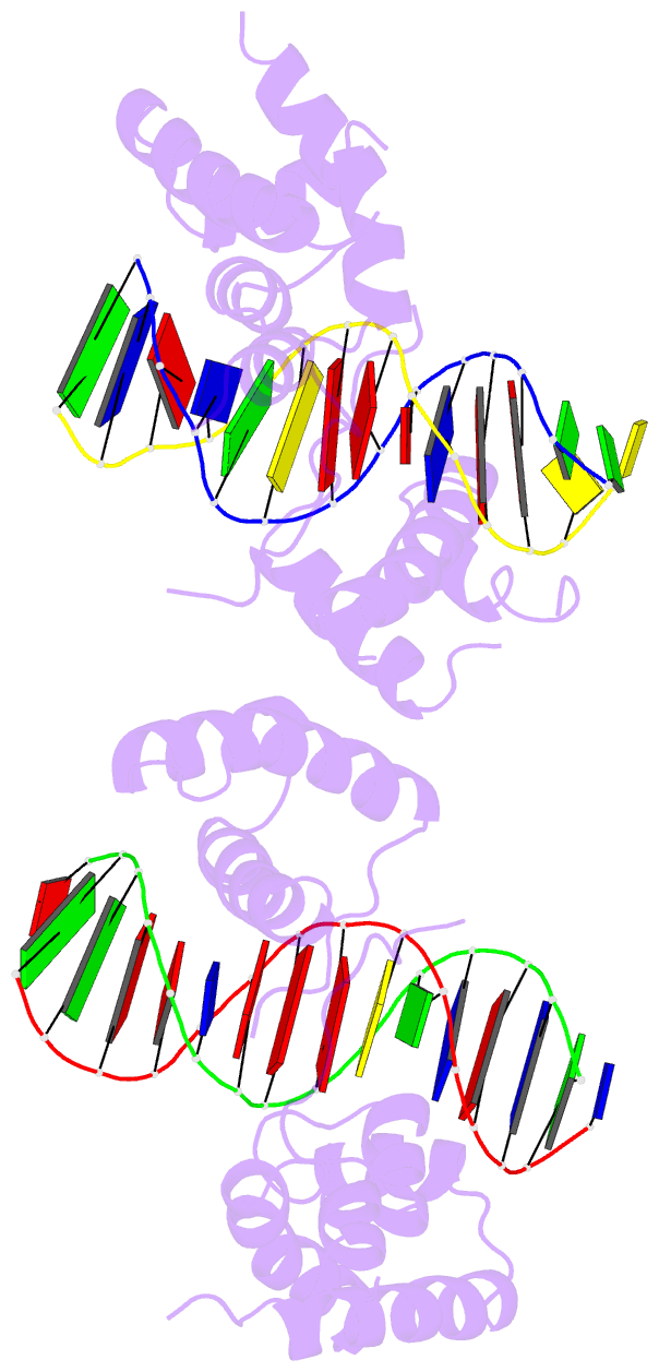
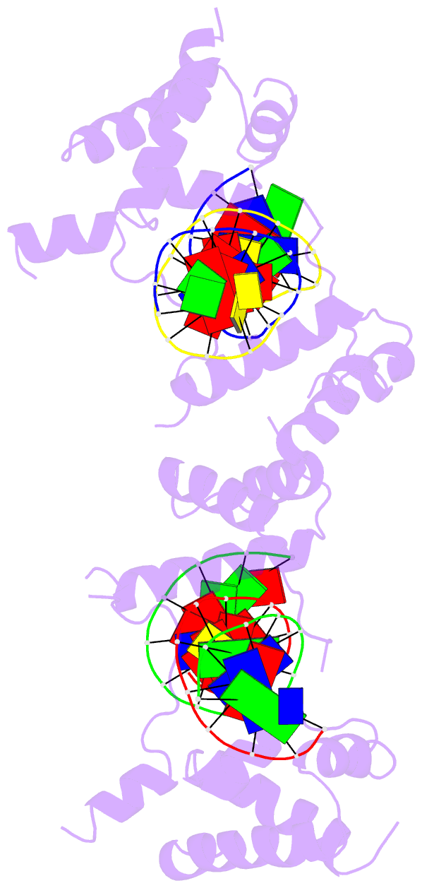
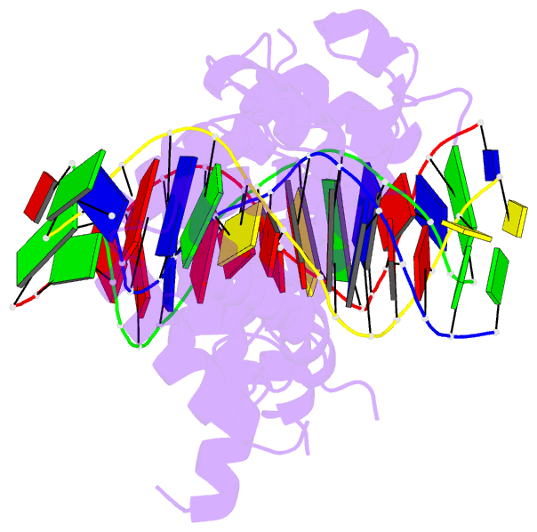
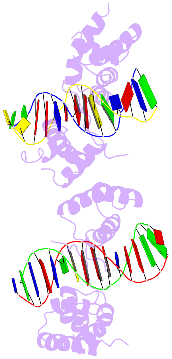
- Crystal structure of a ternary complex containing an oca-b peptide, the oct-1 pou domain, and an octamer element
- Reference
- Chasman D, Cepek K, Sharp PA, Pabo CO (1999): "Crystal structure of an OCA-B peptide bound to an Oct-1 POU domain/octamer DNA complex: specific recognition of a protein-DNA interface." Genes Dev., 13, 2650-2657. doi: 10.1101/gad.13.20.2650.
- Abstract
- We have determined the crystal structure, at 3.2 A, of a ternary complex containing an OCA-B peptide, the Oct-1 POU domain, and an octamer DNA site. The OCA-B peptide binds in the major groove near the center of the octamer site, and its polypeptide backbone forms a pair of hydrogen bonds with the adenine base at position 5 of the octamer DNA. Numerous protein-protein contacts between the OCA-B peptide and the POU domain are also involved in the ternary complex. In particular, the hydrophobic surface from a short alpha-helix of OCA-B helps to stabilize the complex by binding to a hydrophobic pocket on the POU-specific domain. The structure of this ternary complex is consistent with previous biochemical studies and shows how peptide-DNA and peptide-protein contacts from OCA-B provide structural and functional specificity in the regulation of immunoglobulin transcription.