Summary information and primary citation
- PDB-id
- 1bf5; DSSR-derived features in text and JSON formats
- Class
- gene regulation-DNA
- Method
- X-ray (2.9 Å)
- Summary
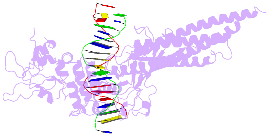
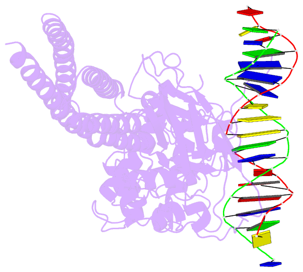
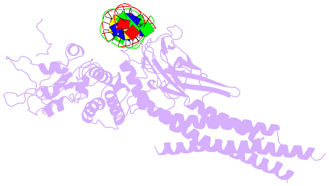
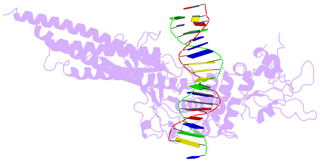
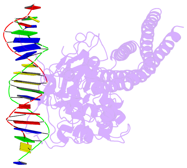
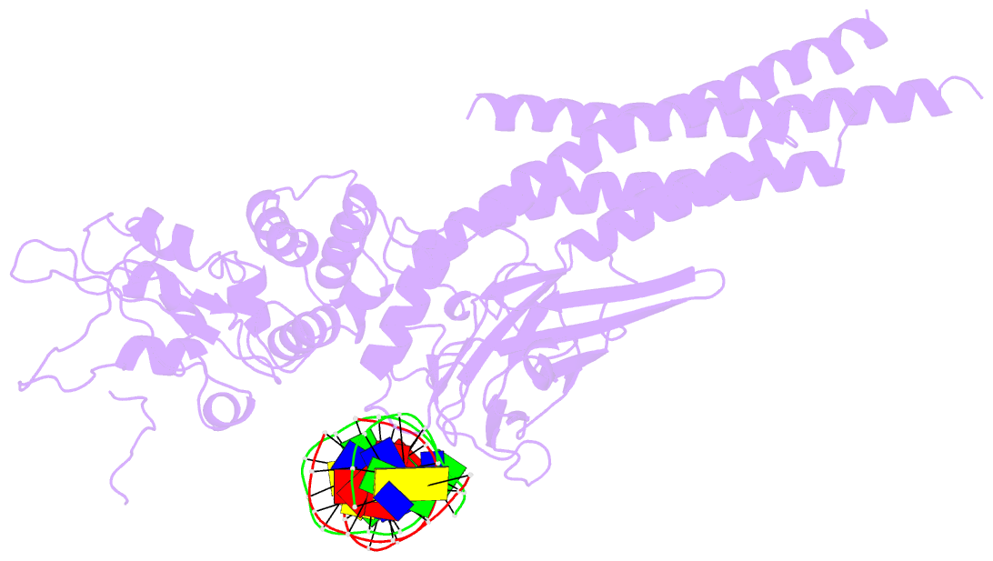
- Tyrosine phosphorylated stat-1-DNA complex
- Reference
- Chen X, Vinkemeier U, Zhao Y, Jeruzalmi D, Darnell Jr JE, Kuriyan J (1998): "Crystal structure of a tyrosine phosphorylated STAT-1 dimer bound to DNA." Cell(Cambridge,Mass.), 93, 827-839. doi: 10.1016/S0092-8674(00)81443-9.
- Abstract
- The crystal structure of the DNA complex of a STAT-1 homodimer has been determined at 2.9 A resolution. STAT-1 utilizes a DNA-binding domain with an immunoglobulin fold, similar to that of NFkappaB and the p53 tumor suppressor protein. The STAT-1 dimer forms a contiguous C-shaped clamp around DNA that is stabilized by reciprocal and highly specific interactions between the SH2 domain of one monomer and the C-terminal segment, phosphorylated on tyrosine, of the other. The phosphotyrosine-binding site of the SH2 domain in each monomer is coupled structurally to the DNA-binding domain, suggesting a potential role for the SH2-phosphotyrosine interaction in the stabilization of DNA interacting elements.