Summary information and primary citation
- PDB-id
- 1an2; DSSR-derived features in text and JSON formats
- Class
- transcription-DNA
- Method
- X-ray (2.9 Å)
- Summary
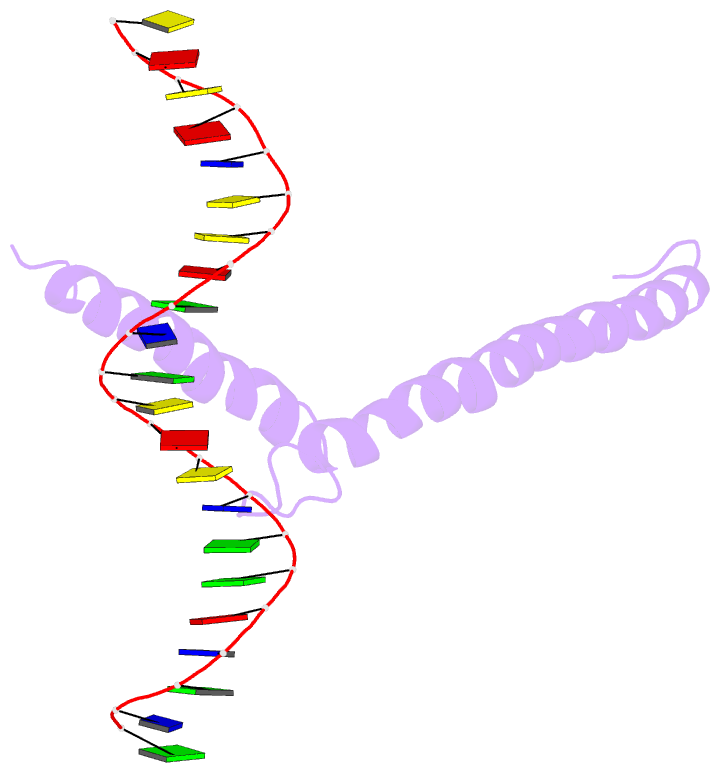
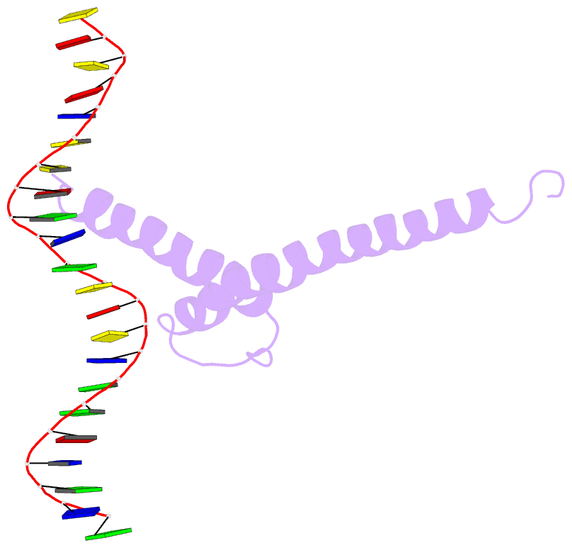
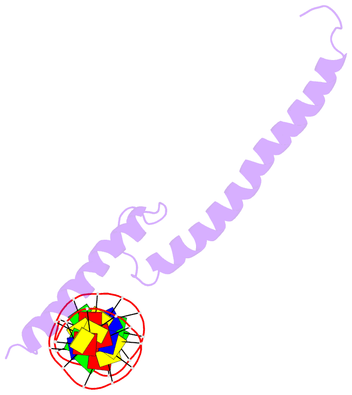
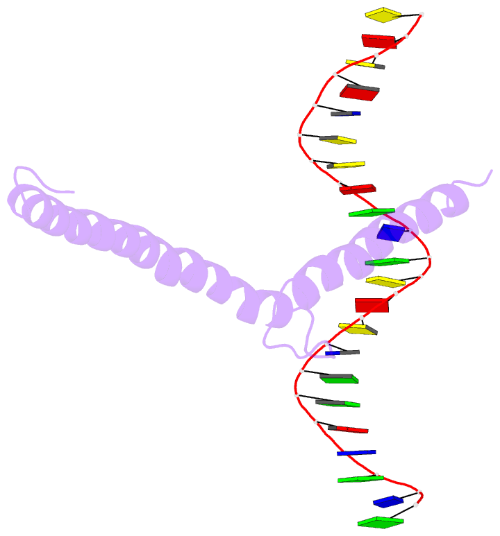
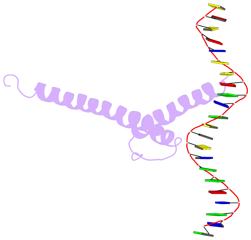
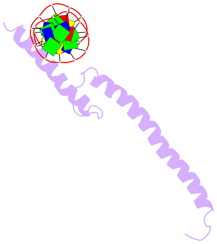
- Recognition by max of its cognate DNA through a dimeric b-hlh-z domain
- Reference
- Ferre-D'Amare AR, Prendergast GC, Ziff EB, Burley SK (1993): "Recognition by Max of its cognate DNA through a dimeric b/HLH/Z domain." Nature, 363, 38-45. doi: 10.1038/363038a0.
- Abstract
- The three-dimensional structure of the basic/helix-loop-helix/leucine zipper domain of the transcription factor Max complexed with DNA has been determined by X-ray crystallography at 2.9 A resolution. Max binds as a dimer to its recognition sequence CACGTG by direct contacts between the alpha-helical basic region and the major groove. This symmetric homodimer, a new protein fold, is a parallel, left-handed, four-helix bundle, with each monomer containing two alpha-helical segments separated by a loop. The two alpha-helical segments are composed of the basic region plus helix 1 and helix 2 plus the leucine repeat, respectively. As in GCN4, the leucine repeat forms a parallel coiled coil.