Summary information and primary citation
- PDB-id
-
166d;
SNAP-derived features in text and
JSON formats
- Class
- DNA
- Method
- X-ray (2.2 Å)
- Summary
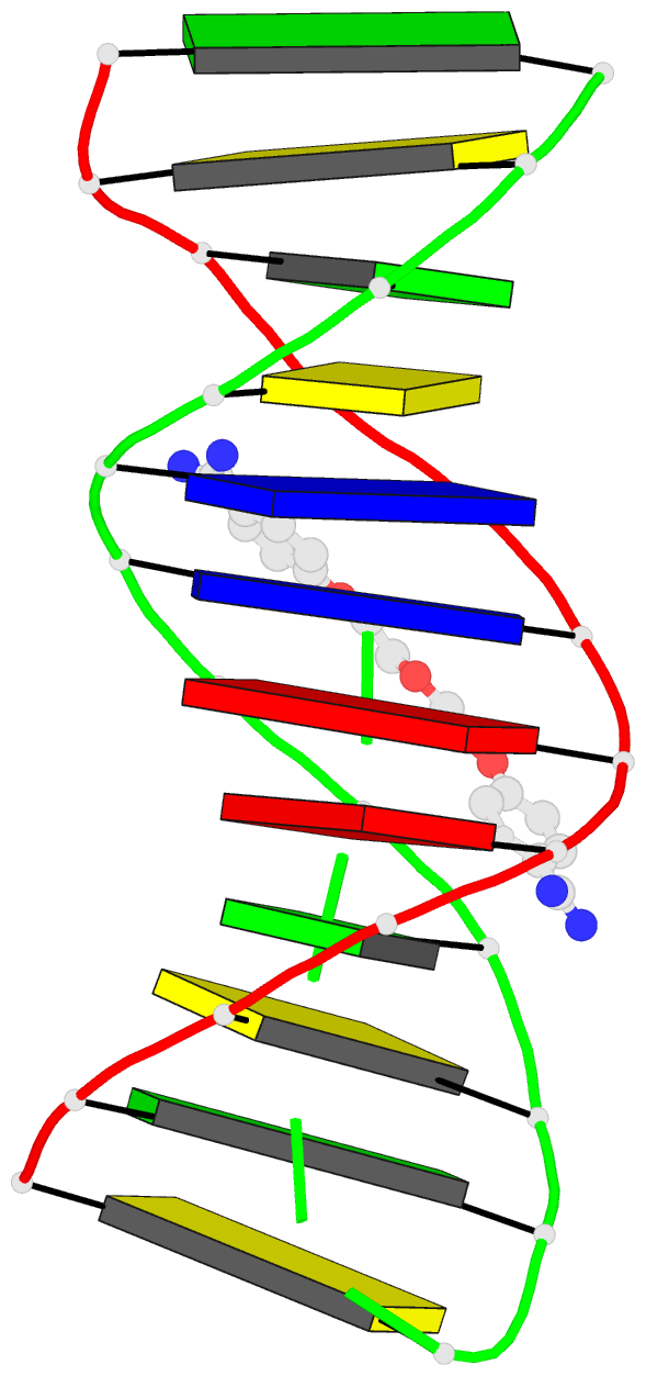
- Drug-DNA minor groove recognition: crystal structure of
gamma-oxapentamidine complexed with d(cgcgaattcgcg)2
- Reference
-
Nunn CM, Jenkins TC, Neidle S (1994): "Crystal
structure of gamma-oxapentamidine complexed with
d(CGCGAATTCGCG)2. The effects of drug structural change
on DNA minor-groove recognition."
Eur.J.Biochem., 226, 953-961.
doi: 10.1111/j.1432-1033.1994.00953.x.
- Abstract
- The crystal structure of the complex of
gamma-oxapentamidine and the DNA dodecamer d(CGCGAATTCGCG)2
has been determined to a resolution of 0.22 nm and an R
factor of 18.9%. The gamma-oxapentamidine ligand interacts
with the dodecamer by classic minor groove binding via
interactions within the A+T-rich region of the minor
groove. A chain of solvent molecules lies along the mouth
of the minor groove on the outside of the bound ligand. The
structural details of the complex are discussed and
compared with the closely analogous complex with
pentamidine bound to the same dodecamer [Edwards, K. J.,
Jenkins, T. C. & Neidle, S. (1992) Biochemistry 31,
7104-7109]. The amidinium groups of the ligand do not
hydrogen bond to bases, but are in close contact with O4'
sugar ring atoms. This in part explains the reduced DNA
binding affinity of this ligand compared to
pentamidine.


