Summary information and primary citation
- PDB-id
-
113d;
SNAP-derived features in text and
JSON formats
- Class
- DNA
- Method
- X-ray (2.5 Å)
- Summary
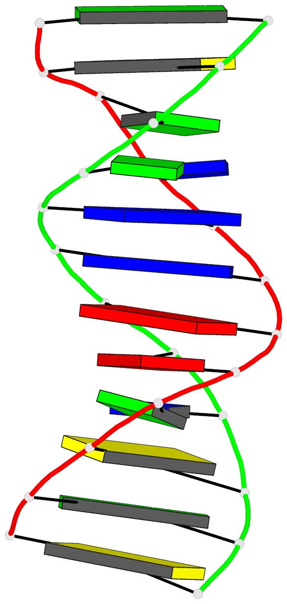
- The structure of guanosine-thymidine mismatches in
b-DNA at 2.5 angstroms resolution
- Reference
-
Hunter WN, Brown T, Kneale G, Anand NN, Rabinovich D,
Kennard O (1987): "The
structure of guanosine-thymidine mismatches in B-DNA at
2.5-A resolution." J.Biol.Chem.,
262, 9962-9970.
- Abstract
- The structure of the deoxyoligomer
d(C-G-C-G-A-A-T-T-T-G-C-G) was determined at 2.5-A
resolution by single crystal x-ray diffraction techniques.
The final R factor is 18% with the location of 71 water
molecules. The oligomer crystallizes in a B-DNA-type
conformation, with two strands interacting to form a
dodecamer duplex. The double helix consists of four A X T
and six G X C Watson-Crick base pairs and two G X T
mismatches. The G X T pairs adopt a "wobble" structure with
the thymine projecting into the major groove and the
guanine into the minor groove. The mispairs are
accommodated in the normal double helix by small
adjustments in the conformation of the sugar phosphate
backbone. A comparison with the isomorphous parent compound
containing only Watson-Crick base pairs shows that any
changes in the structure induced by the presence of G X T
mispairs are highly localized. The global conformation of
the duplex is conserved. The G X T mismatch has already
been studied by x-ray techniques in A and Z helices where
similar results were found. The geometry of the mispair is
essentially identical in all structures so far examined,
irrespective of the DNA conformation. The hydration is also
similar with solvent molecules bridging the functional
groups of the bases via hydrogen bonds. Hydration may be an
important factor in stabilizing G X T mismatches. A
characteristic of Watson-Crick paired A X T and G X C bases
is the pseudo 2-fold symmetry axis in the plane of the base
pairs. The G X T wobble base pair is pronouncedly
asymmetric. This asymmetry, coupled with the disposition of
functional groups in the major and minor grooves, provides
a number of features which may contribute to the
recognition of the mismatch by repair enzymes.


